Trabecular Pattern
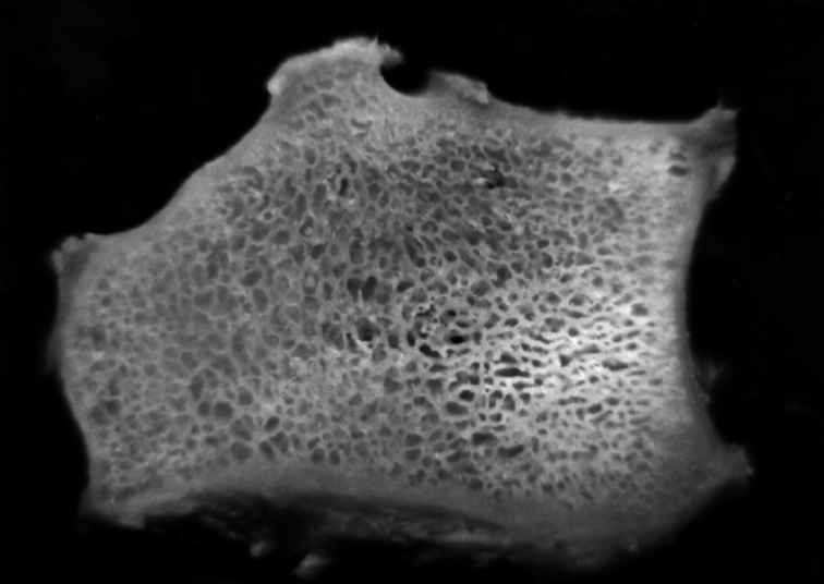
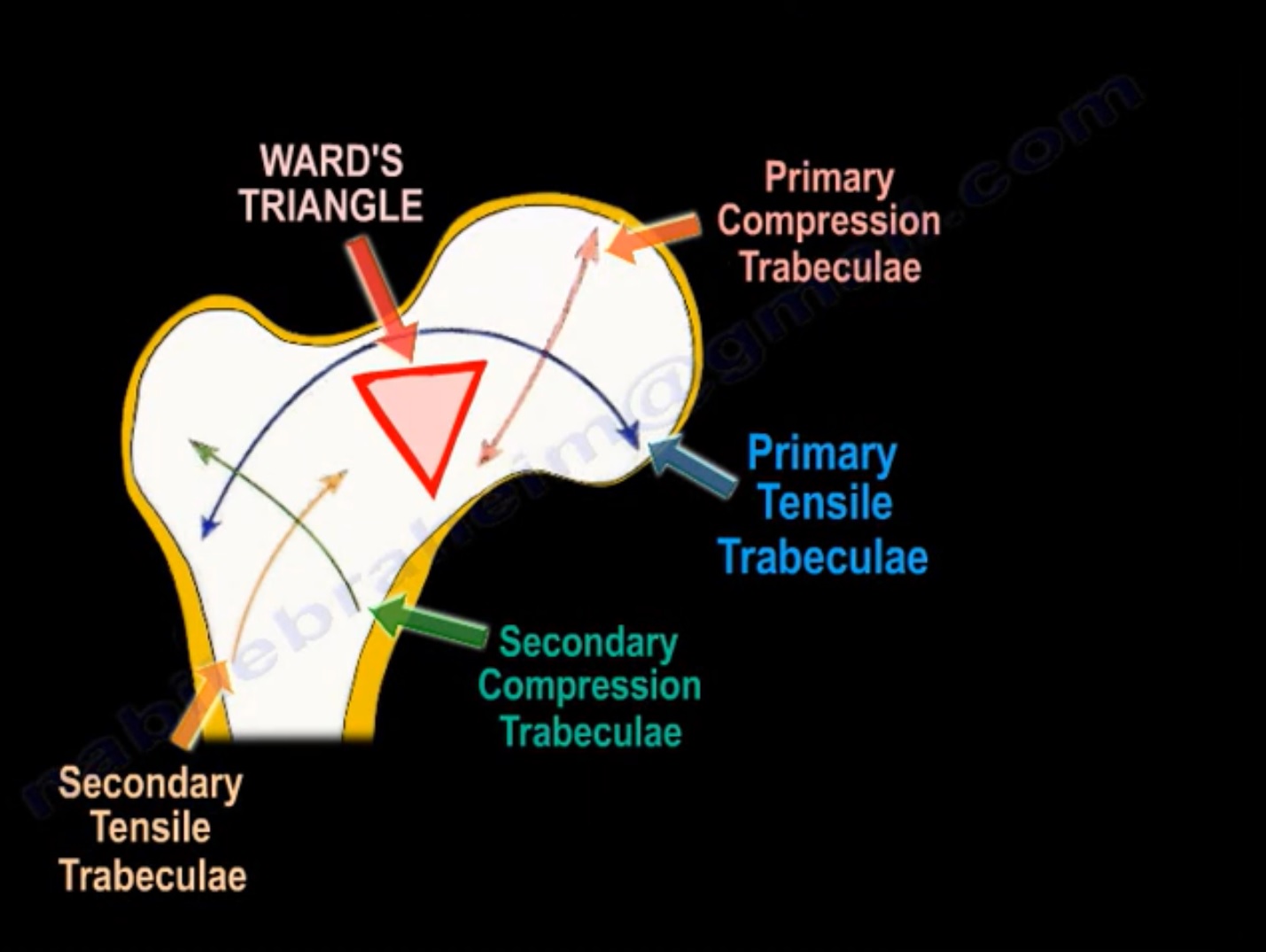
Trabecular Pattern - Web trabecular pattern (concept id: 2000 ), and the distal aspect of the appendicular long bones, particularly the proximal end of femur and the distal end of radius. Harry benjamin laing mrcs, ortho m8, frcs(tr and orth) tutorials. A mnemonic to remember differentials causing a coarse trabecular pattern in the bone. Periapical radiographs of patients who had full mouth intraoral radiographs were collected. Cclinical test, rresearch test, oomim, ggenereviews, vclinvar. Figures 2 and 3 show photos of bone slices of someone with and someone without osteopenia (the thinning of bone that leads to osteoporosis). Citation, doi, disclosures and article data. Web revealing the underlying rule. Neoplasm of follicular cells showing a trabecular growth pattern of large cells with pale to eosinophilic cytoplasm containing stromal hyaline material; Periapical radiographs of patients who had full mouth intraoral radiographs were collected. Web a trabecula ( pl.: Trabeculae, from latin for 'small beam') is a small, often microscopic, tissue element in the form of a small beam, strut or rod that supports or anchors a framework of parts within a body or organ. Web these trabecular patterns are diagramatically represented. Prof nabil ebraheim, university of toledo, ohio, usa. The nuclear features show elongation, grooves and intranuclear inclusions ( am j surg pathol 1987;11:583 ) essential features. Web the trabecular pattern in the upper end of femur is analyzed on the basis of the presence or absence, the relative number and density of the trabeculae, trabecular group and also by the. A microscopic finding indicating that the neoplastic cells are arranged in trabeculae in a tumor sample. Trabeculae groups of proximal femur. Together these trabeculae create the ward triangle. Creep and cyclic loading tests are implemented to model daily mechanical loadings on trabecular bone. Web last revised by owen kang on 26 apr 2020. Citation, doi, disclosures and article data. A square is first divided into two. Web trabecular bone shows the classical creep characteristics with three phases: This study was performed to evaluate possible variations in maxillary and mandibular bone texture of normal population using the fractal analysis, particles count, and area fraction in intraoral radiographs. A microscopic finding indicating that the neoplastic. Marginal bone loss (mbl) is one of the leading causes of dental implant failure. Web trabecular pattern (concept id: Cclinical test, rresearch test, oomim, ggenereviews, vclinvar. Figures 2 and 3 show photos of bone slices of someone with and someone without osteopenia (the thinning of bone that leads to osteoporosis). A square is first divided into two. Hughes, stephan joseph, eric j. Coarse trabecular pattern in bone (mnemonic) A square is first divided into two. Web last revised by owen kang on 26 apr 2020. Cclinical test, rresearch test, oomim, ggenereviews, vclinvar. Web last revised by owen kang on 26 apr 2020. Web the trabecular pattern on panoramic radiographs provides a strong predictor of fractures, at least for postmenopausal women. A microscopic finding indicating that the neoplastic cells are arranged in trabeculae in a tumor sample. Web trabecular pattern of proximal femur refers to the five groups of trabeculae that are demonstrable. High elastic strain response, steady state response, and necking (which the strain rate exponentially increases). Web these trabecular patterns are diagramatically represented in fig. Web last revised by owen kang on 26 apr 2020. Web trabecular pattern of the proximal femur. Web a trabecula ( pl.: Web last revised by daniel j bell on 27 may 2021. You can see the trabecular network directly by looking at vertical slices through two bones; Periapical radiographs of patients who had full mouth intraoral radiographs were collected. Web trabecular bone shows the classical creep characteristics with three phases: High elastic strain response, steady state response, and necking (which the. Trabeculae groups of proximal femur. The assessment by an observer combined with texture analysis procedures yields a predictive power that. The nuclear features show elongation, grooves and intranuclear inclusions ( am j surg pathol 1987;11:583 ) essential features. The disparity of the radiographic images of the slices compared with that of the whole bone suggests that the trabecular pattern as. You can see the trabecular network directly by looking at vertical slices through two bones; Cclinical test, rresearch test, oomim, ggenereviews, vclinvar. Prof nabil ebraheim, university of toledo, ohio, usa. Trabeculae, from latin for 'small beam') is a small, often microscopic, tissue element in the form of a small beam, strut or rod that supports or anchors a framework of parts within a body or organ. High elastic strain response, steady state response, and necking (which the strain rate exponentially increases). Web coarse trabecular bones can result from a number of causes 1,2: Creep and cyclic loading tests are implemented to model daily mechanical loadings on trabecular bone. Coarse trabecular pattern in bone (mnemonic) Citation, doi, disclosures and article data. Marginal bone loss (mbl) is one of the leading causes of dental implant failure. The nuclear features show elongation, grooves and intranuclear inclusions ( am j surg pathol 1987;11:583 ) essential features. Web these trabecular patterns are diagramatically represented in fig. Trabeculae groups of proximal femur. Together these trabeculae create the ward triangle. He explains, take type 1 as an example; Web trabecular pattern of proximal femur refers to the five groups of trabeculae that are demonstrable within the femoral head and neck.
The Trabecular Bone Pattern as a window into Osteoporosis

Trabecular bone patterns Download Scientific Diagram

Trabecular bone Definition of Trabecular bone

Trabecular Bone Images Galleries With A Bite!

Crosssectional images of a trabecular pattern, from different CBCT

Trabecular Pattern of the Proximal Femur —

Typical carcinoid, trabecular pattern Trabeculae are separ… Flickr

Trabecular bone patterns Download Scientific Diagram

Crosssection of human femur showing trabecular and cortical bone from

Scheme of changes in trabecular pattern of the proximal Download
The Disparity Of The Radiographic Images Of The Slices Compared With That Of The Whole Bone Suggests That The Trabecular Pattern As Seen On A Radiograph Of A Bone Could Be The Result Of:
Web The Trabecular Pattern On Panoramic Radiographs Provides A Strong Predictor Of Fractures, At Least For Postmenopausal Women.
Web Last Revised By Daniel J Bell On 27 May 2021.
Thalassemia, Chronic Iron Deficiency Anemia 3;
Related Post: