Teg Interpretation Chart
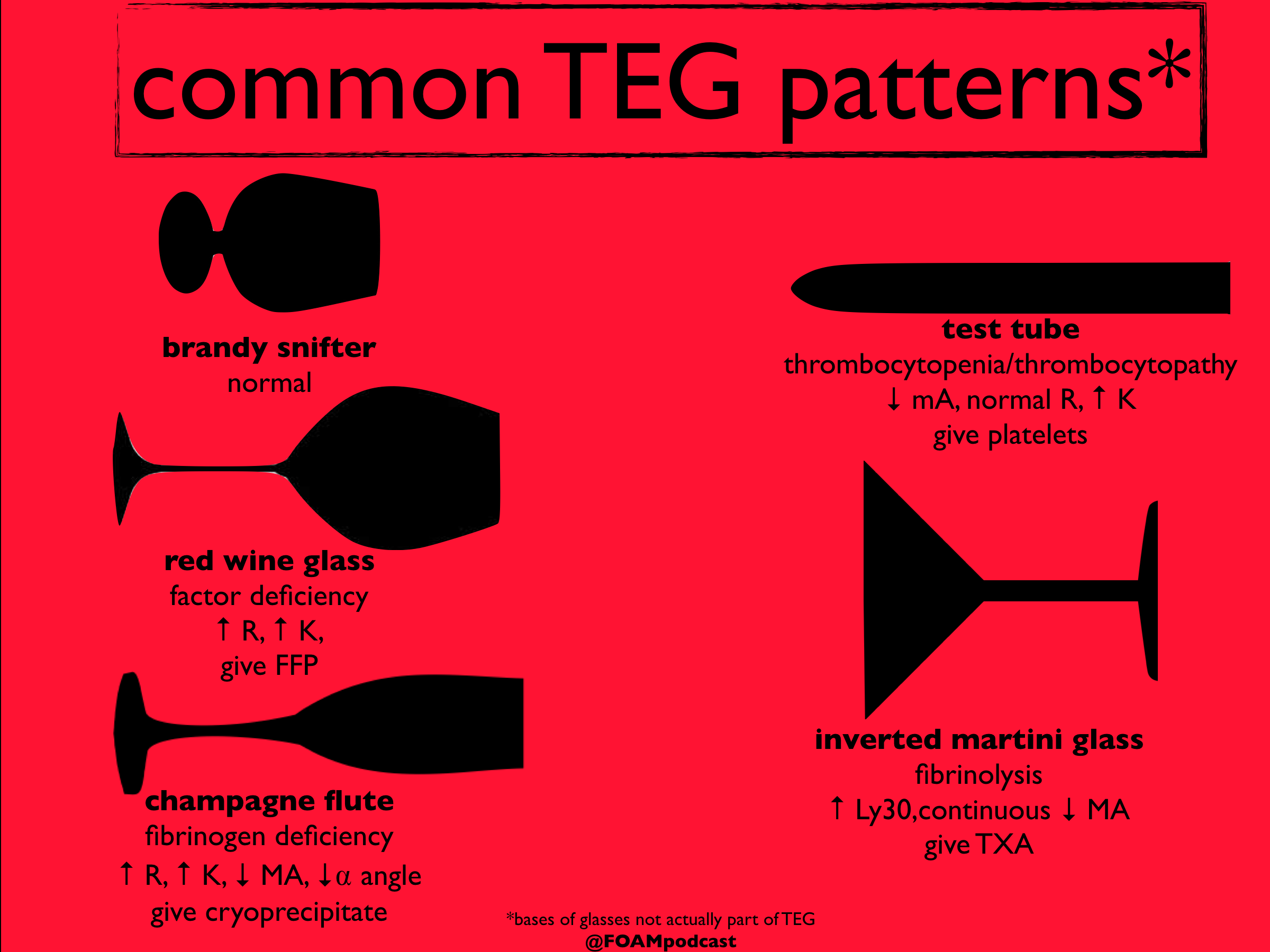
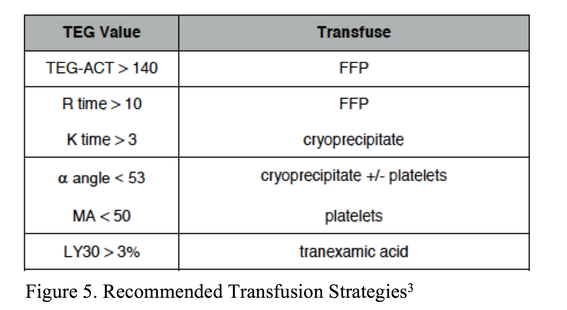
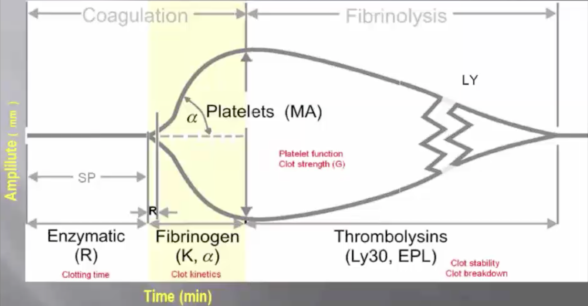
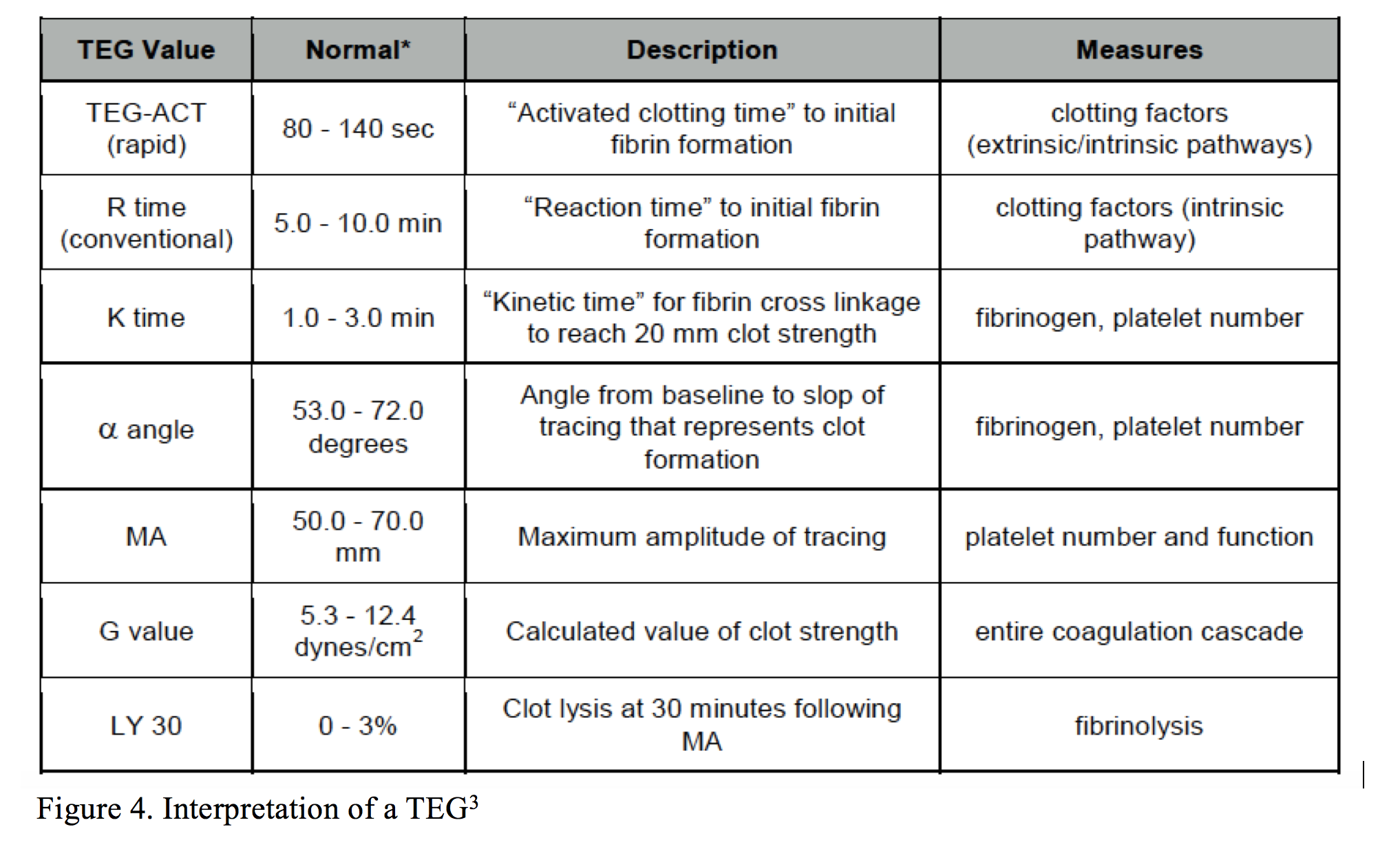
Teg Interpretation Chart - Web a basic guide to assays and clinical interpretation. The teg analyzer consists of an electronic recorder attached to a torsion wire that suspends a plastic pin into a plastic cup or cuvette. It is a test mainly used in surgery and anesthesiology, although increasingly used in resuscitations in emergency departments,. Teg 5000 (units) rotem delta (units) reaction rate (r) (min) clotting time (ct) (s) time for the trace to reach an amplitude of 2 mm. Replace them with fresh frozen plasma (ffp). Thromboelastography (teg) is a valuable diagnostic tool that assesses blood clotting ability, providing a comprehensive evaluation of the coagulation system and aiding in the identification of abnormalities in blood clotting. Web as the blood begins to clot and adhere to the pin, the movement of the pin increases. Em, depicts a normal teg. Rotem® results in clinically abnormal temograms significant bleeding. Teg can help to better identify coagulopathy and. Web teg ® is a different test in that it measures the viscoelastic properties of whole blood as it clots. Web as seen in figure 2, an r time of 1.8min indicates a fairly significant hypercoagulable state, which required treatment with full dose heparin anticoagulation for treatment of an acute deep vein thrombosis, despite an inr of 4. Replace them. Teg can identify patients who have coagulopathy and can guide blood product transfusions. Web thromboelastography (teg) is a laboratory assay that measures the viscoelastic properties of whole blood clotting. Teg 5000 (units) rotem delta (units) reaction rate (r) (min) clotting time (ct) (s) time for the trace to reach an amplitude of 2 mm. Thromboelastography is used to identify acute. Rotem® results in clinically abnormal temograms significant bleeding. This increasing movement is interpreted by the computer as increasing amplitude on the teg graph. Web so, how do we interpret tegs? Web interpretation of teg factsheet v1 final march 2013.doc page 2 of 2 clot stability (ly30/epl)/maximum lysis (ml) once the clot achieves its maximum clot strength it then begins to. If the r time is elevated, this implies a deficiency in clotting factors. This increasing movement is interpreted by the computer as increasing amplitude on the teg graph. Web thromboelastography (teg) is a laboratory assay that measures the viscoelastic properties of whole blood clotting. Web a basic guide to assays and clinical interpretation. Web this guideline reviews the interpretation of. The teg analyzer consists of an electronic recorder attached to a torsion wire that suspends a plastic pin into a plastic cup or cuvette. Time elapsed till clot initially forms. (demonstrating coagulopathies) (ct, a10 (mcf) and ml): Web teg ® is a different test in that it measures the viscoelastic properties of whole blood as it clots. If the r. Thromboelastography ( teg) is a method of testing the efficiency of blood coagulation. The teg analyzer consists of an electronic recorder attached to a torsion wire that suspends a plastic pin into a plastic cup or cuvette. (demonstrating coagulopathies) (ct, a10 (mcf) and ml): Teg with citrated kaolin activator. In this chapter, we very briefly review teg applications and discuss. Web thromboelastography (teg) is a laboratory assay that measures the viscoelastic properties of whole blood clotting. Web interpretation of teg factsheet v1 final march 2013.doc page 2 of 2 clot stability (ly30/epl)/maximum lysis (ml) once the clot achieves its maximum clot strength it then begins to break down over a few hours. Web this guideline reviews the interpretation of a. Thromboelastography assesses the mechanics of clot formation and fibrinolysis through a pin suspended in a blood sample. If the r time is elevated, this implies a deficiency in clotting factors. Web this guideline reviews the interpretation of a thromboelastogram and how it may be used to guide blood product administration. Web as seen in figure 2, an r time of. It provides global information on the dynamics of clot development, stabilization and dissolution which reflects in vivo hemostasis. It is a test mainly used in surgery and anesthesiology, although increasingly used in resuscitations in emergency departments,. Teg can identify patients who have coagulopathy and can guide blood product transfusions. Web interpretation of teg factsheet v1 final march 2013.doc page 2. Web as the blood begins to clot and adhere to the pin, the movement of the pin increases. It provides global information on the dynamics of clot development, stabilization and dissolution which reflects in vivo hemostasis. Web as seen in figure 2, an r time of 1.8min indicates a fairly significant hypercoagulable state, which required treatment with full dose heparin. Teg can help to better identify coagulopathy and. Thromboelastography assesses the mechanics of clot formation and fibrinolysis through a pin suspended in a blood sample. If the r time is elevated, this implies a deficiency in clotting factors. The image below, courtesy of r.e.b.e.l. In this chapter, we very briefly review teg applications and discuss interpretations, normal ranges, and reference controls, and we explain the method of. Thromboelastography ( teg) is a method of testing the efficiency of blood coagulation. Web current teg assays use a variety of samples and can vary slightly in the procedures. Em, depicts a normal teg. Web • teg may be used to guide blood product administration in bleeding patients as follows: Teg with citrated kaolin activator. Web thromboelastography (teg) is a laboratory assay that measures the viscoelastic properties of whole blood clotting. The degree to which this occurs is measured by. It is a test mainly used in surgery and anesthesiology, although increasingly used in resuscitations in emergency departments,. Thromboelastography is used to identify acute coagulopathies in both. Thromboelastography (teg) is a valuable diagnostic tool that assesses blood clotting ability, providing a comprehensive evaluation of the coagulation system and aiding in the identification of abnormalities in blood clotting. Web a basic guide to assays and clinical interpretation.
THROMBOELASTOGRAPHY (TEG) General Principle • A small GrepMed

Thromboelastography (TEG) Guided Resuscitation FOAMcast

Thromboelastography (trademarked as TEG) • Graphical GrepMed

Emergency Medicine EducationThe Thromboelastogram (TEG

The Use of TEG & Goal Directed Blood Component Therapy University of

REBEL Review 54 Thromboelastogram (TEG) Interpretation R GrepMed

Emergency Medicine EducationThe Thromboelastogram (TEG

Thromboelastography aka The TEG — Taming the SRU

Thromboelastography in Pediatric Critical Care PPAG News

TEG Cheat Sheet
This Increasing Movement Is Interpreted By The Computer As Increasing Amplitude On The Teg Graph.
Web As Seen In Figure 2, An R Time Of 1.8Min Indicates A Fairly Significant Hypercoagulable State, Which Required Treatment With Full Dose Heparin Anticoagulation For Treatment Of An Acute Deep Vein Thrombosis, Despite An Inr Of 4.
It Provides Global Information On The Dynamics Of Clot Development, Stabilization And Dissolution Which Reflects In Vivo Hemostasis.
Printed With Permission Of 2022 Augusta University And Peter Naktin.
Related Post: