Strain Pattern In Ecg
Strain Pattern In Ecg - Such hypertrophy is usually the. Web the most commonly observed pattern is asymmetrical thickening of the anterior interventricular septum (= asymmetrical septal hypertrophy ). Asymptomatic patients with nf1 had normal. Web left ventricular hypertrophy with strain pattern ecg (example 1) | learn the heart. Web lvh with strain pattern can sometimes be seen in long standing severe aortic regurgitation, usually with associated left ventricular hypertrophy and systolic. Web electrocardiographic strain pattern is associated with left ventricular concentric remodeling, scar, and mortality over 10 years: Web an ecg strain pattern was present in 101 patients (23%). Changes need to occur in at least 2 of the right. Web left ventricular hypertrophy with strain pattern (example 3) | learn the heart. Web a study in the american journal of cardiology published in 2017 showed that ecg strain—found in 28% of patients undergoing surgical aortic valve replacement for. Web left ventricular hypertrophy (lvh) refers to an increase in the size of myocardial fibers in the main cardiac pumping chamber. Its presence on the ecg of hypertensive. Web a study in the american journal of cardiology published in 2017 showed that ecg strain—found in 28% of patients undergoing surgical aortic valve replacement for. This pattern is associated with high.. Changes need to occur in at least 2 of the right. Web left ventricular hypertrophy with strain pattern (example 3) | learn the heart. Web a study in the american journal of cardiology published in 2017 showed that ecg strain—found in 28% of patients undergoing surgical aortic valve replacement for. Web electrocardiographic strain pattern is associated with left ventricular concentric. Such hypertrophy is usually the. Web electrocardiographic strain pattern is associated with left ventricular concentric remodeling, scar, and mortality over 10 years: Changes need to occur in at least 2 of the right. S wave in v2 + r wave in v5 > 35 mm. Web left ventricular hypertrophy (lvh) refers to an increase in the size of myocardial fibers. Web the most commonly observed pattern is asymmetrical thickening of the anterior interventricular septum (= asymmetrical septal hypertrophy ). Such hypertrophy is usually the. Web a study in the american journal of cardiology published in 2017 showed that ecg strain—found in 28% of patients undergoing surgical aortic valve replacement for. Its presence on the ecg of hypertensive. Changes need to. This pattern is associated with high. S wave in v2 + r wave in v5 > 35 mm. Its presence on the ecg of hypertensive. Web an ecg strain pattern was present in 101 patients (23%). Changes need to occur in at least 2 of the right. Web electrocardiographic strain pattern is associated with left ventricular concentric remodeling, scar, and mortality over 10 years: Web left ventricular hypertrophy with strain pattern ecg (example 1) | learn the heart. This pattern is associated with high. Web the most commonly observed pattern is asymmetrical thickening of the anterior interventricular septum (= asymmetrical septal hypertrophy ). Its presence on the. Web lvh with strain pattern can sometimes be seen in long standing severe aortic regurgitation, usually with associated left ventricular hypertrophy and systolic. S wave in v2 + r wave in v5 > 35 mm. Web a study in the american journal of cardiology published in 2017 showed that ecg strain—found in 28% of patients undergoing surgical aortic valve replacement. Changes need to occur in at least 2 of the right. Web left ventricular hypertrophy (lvh) refers to an increase in the size of myocardial fibers in the main cardiac pumping chamber. Such hypertrophy is usually the. This pattern is associated with high. Web electrocardiographic left ventricular hypertrophy with strain pattern has been documented as a marker for left ventricular. Web left ventricular hypertrophy (lvh) refers to an increase in the size of myocardial fibers in the main cardiac pumping chamber. Web electrocardiographic left ventricular hypertrophy with strain pattern has been documented as a marker for left ventricular hypertrophy. Changes need to occur in at least 2 of the right. S wave in v2 + r wave in v5 >. Web left ventricular hypertrophy with strain pattern (example 3) | learn the heart. Web electrocardiographic left ventricular hypertrophy with strain pattern has been documented as a marker for left ventricular hypertrophy. Such hypertrophy is usually the. Changes need to occur in at least 2 of the right. Web lvh with strain pattern can sometimes be seen in long standing severe. Asymptomatic patients with nf1 had normal. S wave in v2 + r wave in v5 > 35 mm. Its presence on the ecg of hypertensive. Web electrocardiographic left ventricular hypertrophy with strain pattern has been documented as a marker for left ventricular hypertrophy. Web lvh with strain pattern can sometimes be seen in long standing severe aortic regurgitation, usually with associated left ventricular hypertrophy and systolic. Web electrocardiographic strain pattern is associated with left ventricular concentric remodeling, scar, and mortality over 10 years: Web there are three ecg patterns associated with brugada syndrome, of which only the type 1 ecg is diagnostic. This pattern is associated with high. Web left ventricular hypertrophy with strain pattern (example 3) | learn the heart. Web the most commonly observed pattern is asymmetrical thickening of the anterior interventricular septum (= asymmetrical septal hypertrophy ). Web left ventricular hypertrophy (lvh) refers to an increase in the size of myocardial fibers in the main cardiac pumping chamber. Web an ecg strain pattern was present in 101 patients (23%). Web left ventricular hypertrophy with strain pattern ecg (example 1) | learn the heart.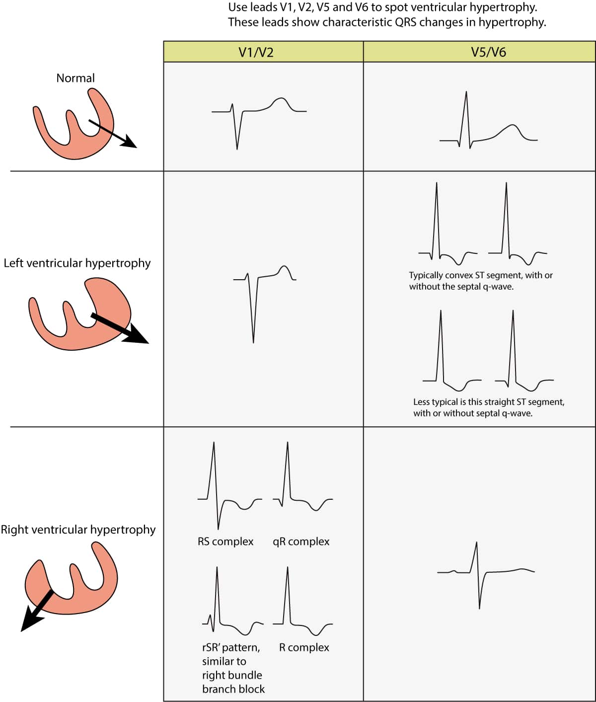
ECG in left ventricular hypertrophy (LVH) criteria and implications
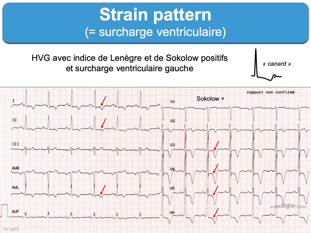
Strain pattern ecardiogram
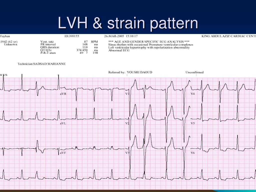
PPT ECG PRACTICAL APPROACH PowerPoint Presentation, free download
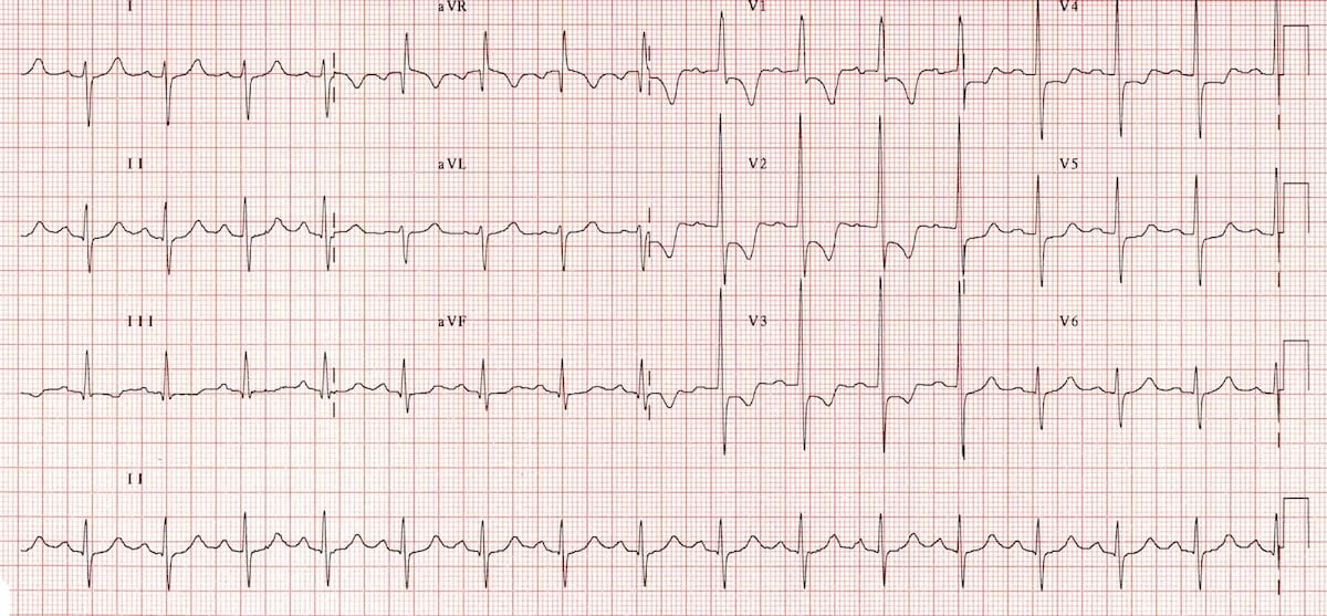
Right Ventricular Strain • LITFL • ECG Library Diagnosis
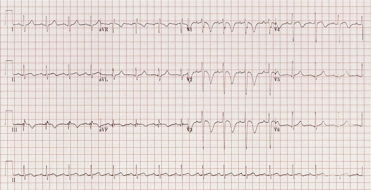
Right Ventricular Strain • LITFL • ECG Library Diagnosis
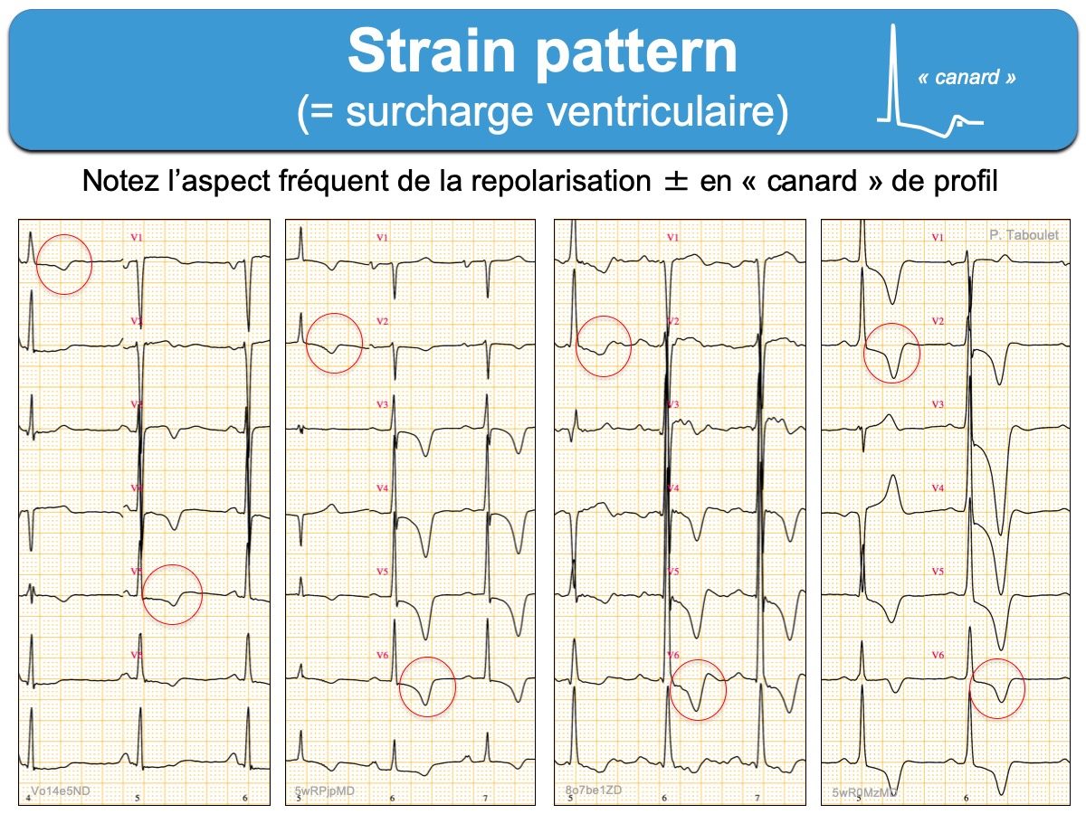
Strain pattern ecardiogram
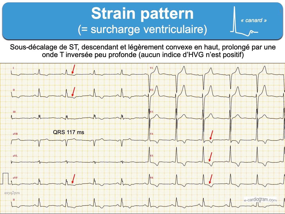
Strain pattern ecardiogram
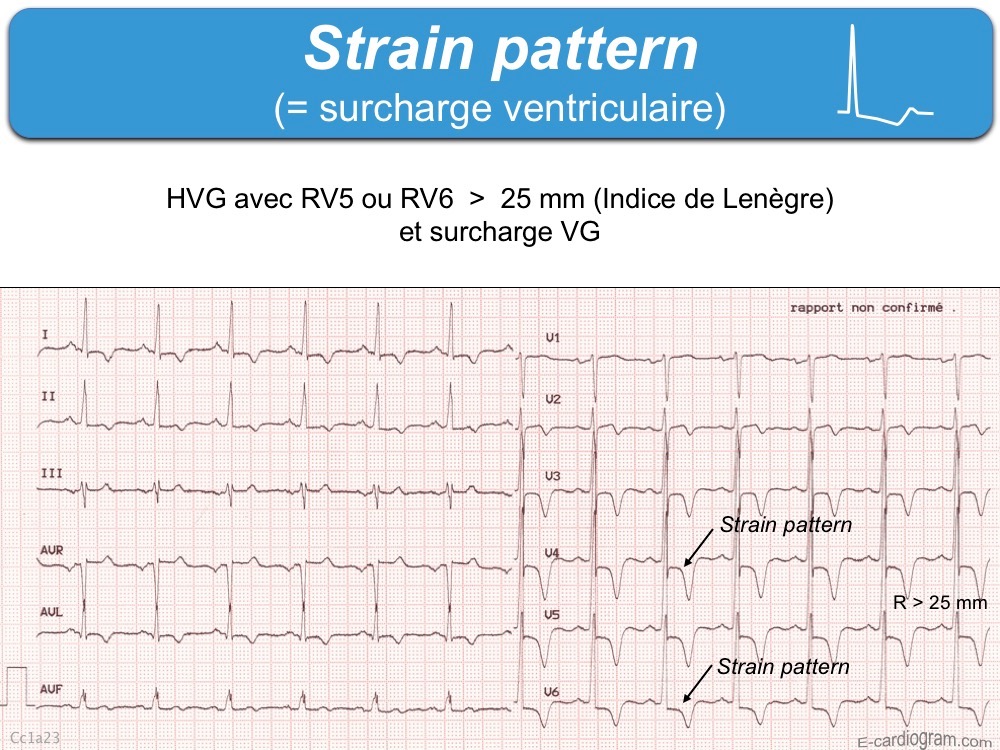
Strain pattern ecardiogram
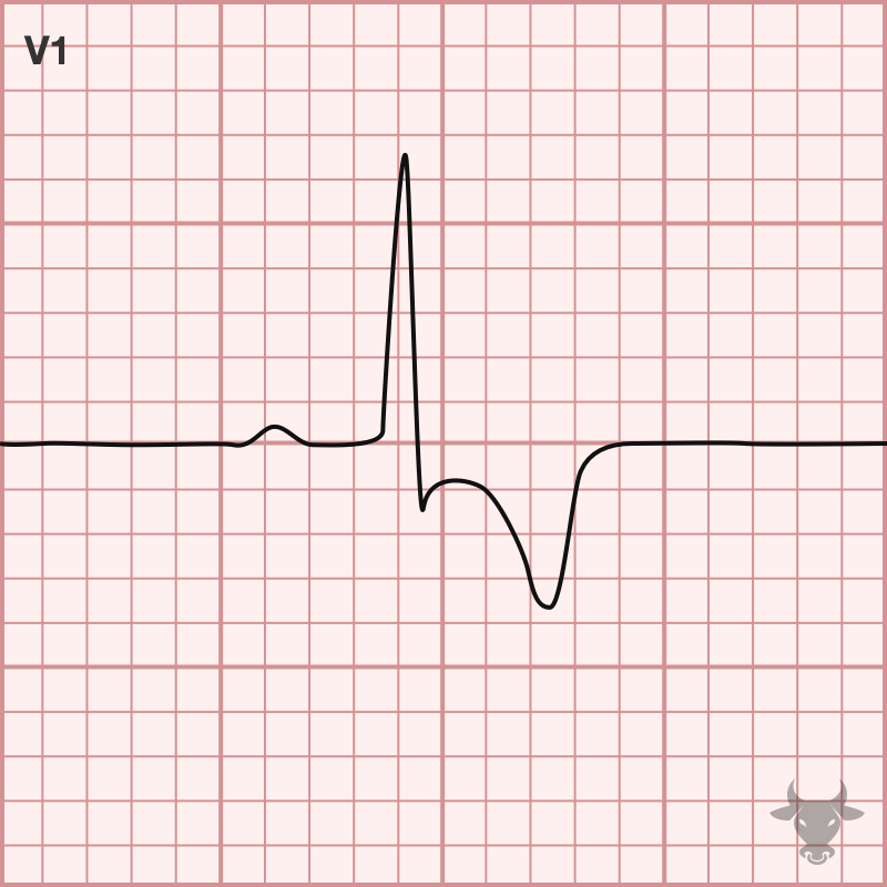
Right Heart Strain ECG Stampede
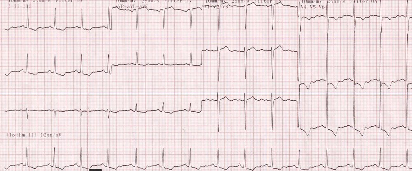
Left ventricular hypertrophy (LVH) with strain pattern
Web A Study In The American Journal Of Cardiology Published In 2017 Showed That Ecg Strain—Found In 28% Of Patients Undergoing Surgical Aortic Valve Replacement For.
Changes Need To Occur In At Least 2 Of The Right.
Ecg Lv Strain Pattern Is Known To Accurately Reflect Replacement Fibrosis In Aortic Stenosis And Hypertensive Cardiomyopathy.
Such Hypertrophy Is Usually The.
Related Post: