Spine Levels Chart
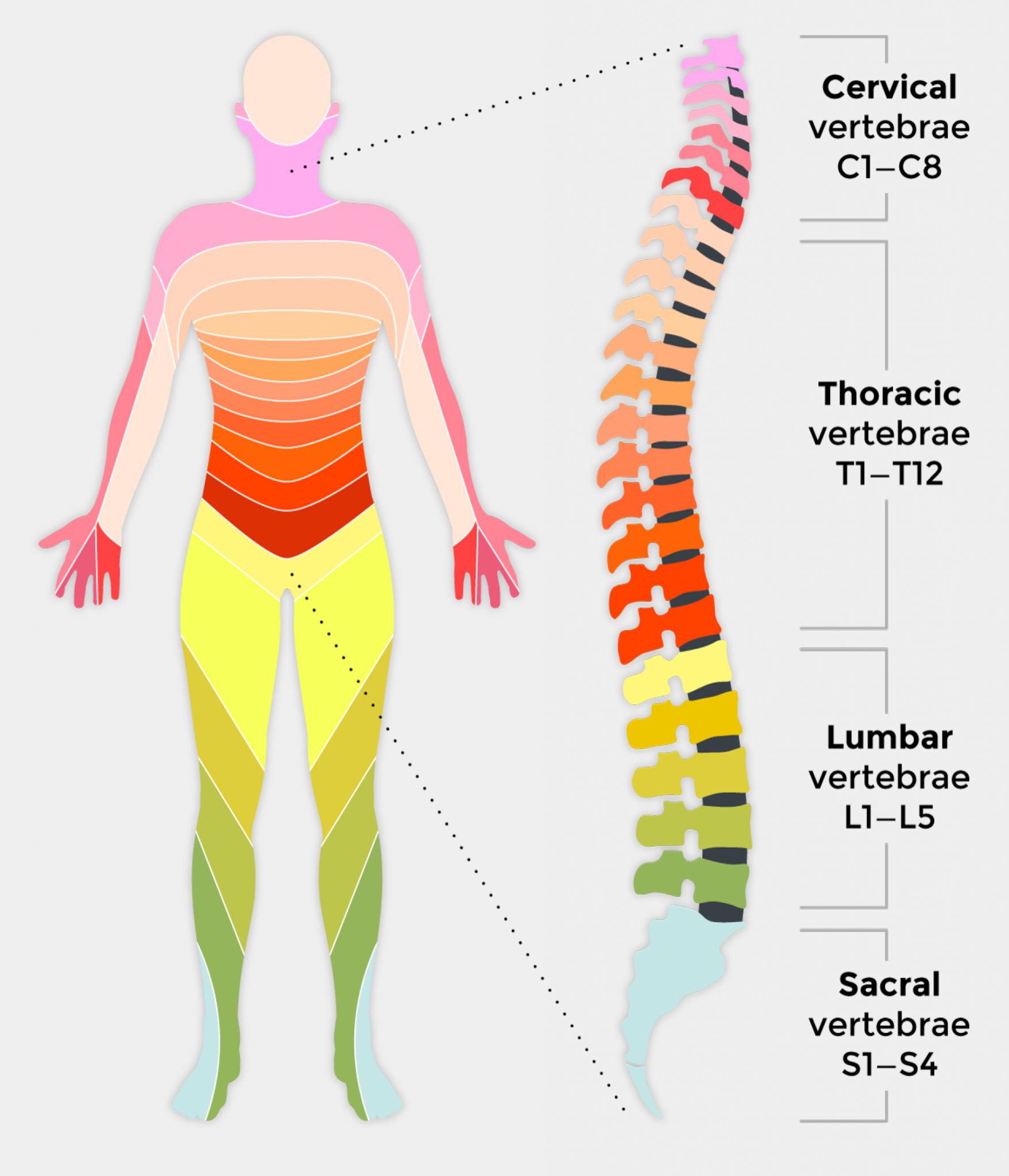
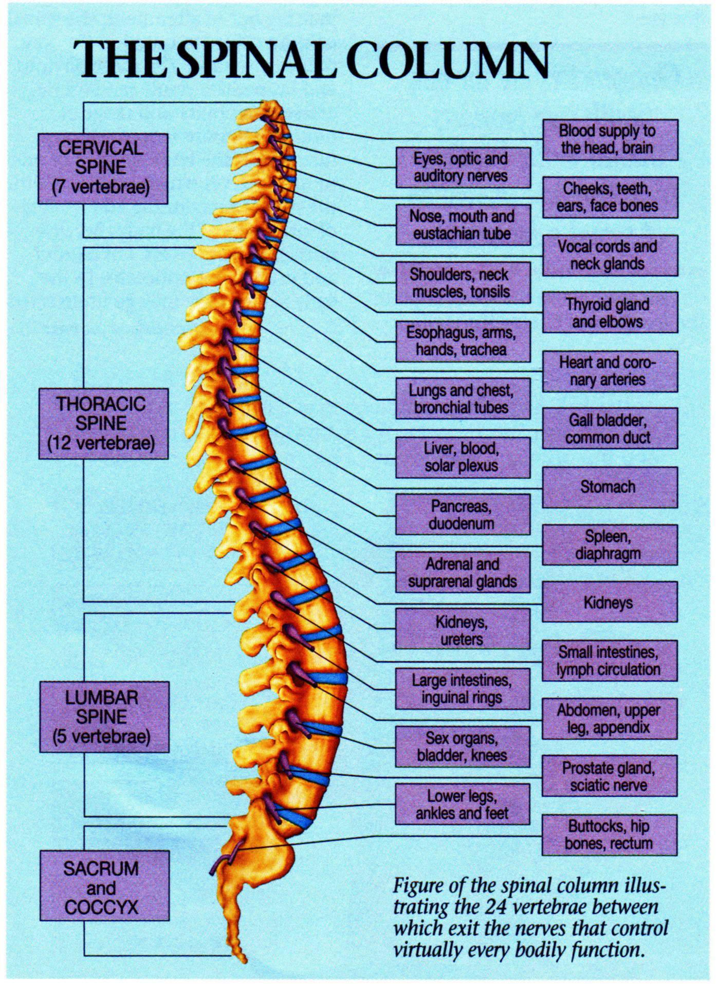
Spine Levels Chart - Web the spine itself has three main segments: Vertebrae are also sometimes called vertebral bodies. The cervical, the thoracic, the lumbar, and the sacral. Vertebral levels are commonly tested in medical examinations. The thoracic is the center portion of the spine, consisting of 12 vertebrae. Your spine starts at the base of your skull (head bone) and ends at your tailbone, a part of your pelvis (the large bony structure between your abdomen and legs). A dermatome is an area of skin supplied by a single spinal nerve. Each vertebra is attached to the one above and below it by ligaments and muscles. Five lumbar spinal nerves on each side called l1 through l5. Web to understand this intricate region, we will consider the bony structures first, and then discuss the ligaments, nerves, and musculature that are associated with this region of the spinal column, concluding with some clinical implications of damage to some of these structures. Each vertebra has specific anatomical features and functions. Cervical (c), thoracic (t), lumbar (l), sacral (s) and coccygeal (cx). Web there are five levels, staring at the top and going downward: The cervical, the thoracic, the lumbar, and the sacral. The thoracic is the center portion of the spine, consisting of 12 vertebrae. Web seven bones in the neck—the cervical spine. The cervical spine, the thoracic spine, and the lumbar spine. It is essential for many functions, such as movement, support, and protecting the spinal. 12 bones in the chest—the thoracic spine. The spine can be divided into five regions: Web seven bones in the neck—the cervical spine. Many of the nerves of the. Cervical (c), thoracic (t), lumbar (l), sacral (s) and coccygeal (cx). Web ‘essential clinical surface anatomy’ is available for purchase here. Web a general view of the spine with the various levels (cervical, thoracic and lumbar spinal regions, sacrum and coccyx) as well as the physiological. 12 bones in the chest—the thoracic spine. Web a general view of the spine with the various levels (cervical, thoracic and lumbar spinal regions, sacrum and coccyx) as well as the physiological curvature of the spine (cervical and lumbar lordosis, thoracic and sacral kyphosis). The back is the body region between the neck and the gluteal regions. It is part. 12 bones in the chest—the thoracic spine. Web a general view of the spine with the various levels (cervical, thoracic and lumbar spinal regions, sacrum and coccyx) as well as the physiological curvature of the spine (cervical and lumbar lordosis, thoracic and sacral kyphosis). Web the human spine consists of 33 vertebrae: The back is the body region between the. Each vertebra has specific anatomical features and functions. Web below is a summary of vertebral levels and associated internal or surface anatomy. Cervical (c), thoracic (t), lumbar (l), sacral (s) and coccygeal (cx). The spine, or backbone, is a long column of bones that runs down the center of a person’s back. Superficial back muscles [17:28] attachments, innervation and functions. Five lumbar spinal nerves on each side called l1 through l5. Web there are five levels, staring at the top and going downward: So, from top to bottom: The spinal cord is the neural passageway that allows for communication between the brain and body. Web the human spine consists of 33 vertebrae: Web how to use the spinal nerve chart: Five bones in the lower back—the lumbar spine. So, from top to bottom: The lower portion of the spine is called the lumbar spine. Web to understand this intricate region, we will consider the bony structures first, and then discuss the ligaments, nerves, and musculature that are associated with this region of. Vertebral levels are commonly tested in medical examinations. Hover over each part to see what they do. The cervical spine, the thoracic spine, and the lumbar spine. The lower portion of the spine is called the lumbar spine. Five lumbar spinal nerves on each side called l1 through l5. Anterior view, posterior view, lateral view , 3d rendering. Five lumbar spinal nerves on each side called l1 through l5. Web below is a summary of vertebral levels and associated internal or surface anatomy. Web to understand this intricate region, we will consider the bony structures first, and then discuss the ligaments, nerves, and musculature that are associated with this. Superficial back muscles [17:28] attachments, innervation and functions of the superficial muscles of the back. The cervical spine, the thoracic spine, and the lumbar spine. A dermatome is an area of skin supplied by a single spinal nerve. Twelve thoracic spinal nerves in each side of the body called t1 through t12. The cervical, the thoracic, the lumbar, and the sacral. They are separated from the vertebra above and below it by an intervertebral. Web the spine diagram below highlights all of the vertebrae labeled. So, from top to bottom: Anterior view, posterior view, lateral view , 3d rendering. Web the human spine consists of 33 vertebrae: The spine, or backbone, is a long column of bones that runs down the center of a person’s back. Web to understand this intricate region, we will consider the bony structures first, and then discuss the ligaments, nerves, and musculature that are associated with this region of the spinal column, concluding with some clinical implications of damage to some of these structures. It is essential for many functions, such as movement, support, and protecting the spinal. These main sections include a group of vertebrae, each of which is given a number that increases as you move down the spine. Web there are 31 pairs of spinal nerves: Hover over each part to see what they do.
10 Facts About Spinal Cord Injury Shield HealthCare

Spinal Anatomy Spinal Regions Bones and Discs Vertebrae Spinal Cord

Mid Back Pain Chiropractor San Diego Dr. Steve Jones Chiropractic

Spinal Cord Levels Chart

What is a Spinal Cord Injury (SCI)? First Aid for Free

A healthy spine is important to your overall health and wellbeing

Spine Chart ANDERSON CHIROPRACTIC

View and Download highresolution Spinal Cord Injury Levels for free

spinal organ chart Premier Chiropractic Centre

Spinal chart You Make it 1Corinthians 620 Pinterest
Five Lumbar Spinal Nerves On Each Side Called L1 Through L5.
Eight Cervical Spinal Nerves On Each Side Of The Spine Called C1 Through C8.
The Spinal Cord Runs Through Its Center.
On The Chart Below You Will See 4 Columns (Vertebral Level, Nerve Root, Innervation, And Possible Symptoms).
Related Post: