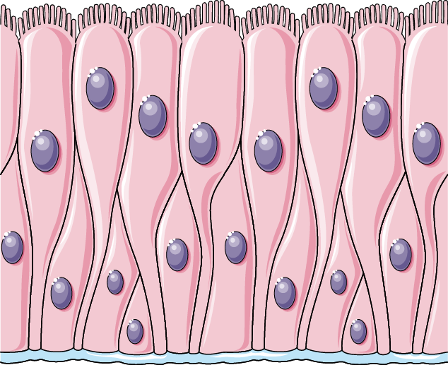
Pseudostratified Ciliated Columnar Epithelium Drawing
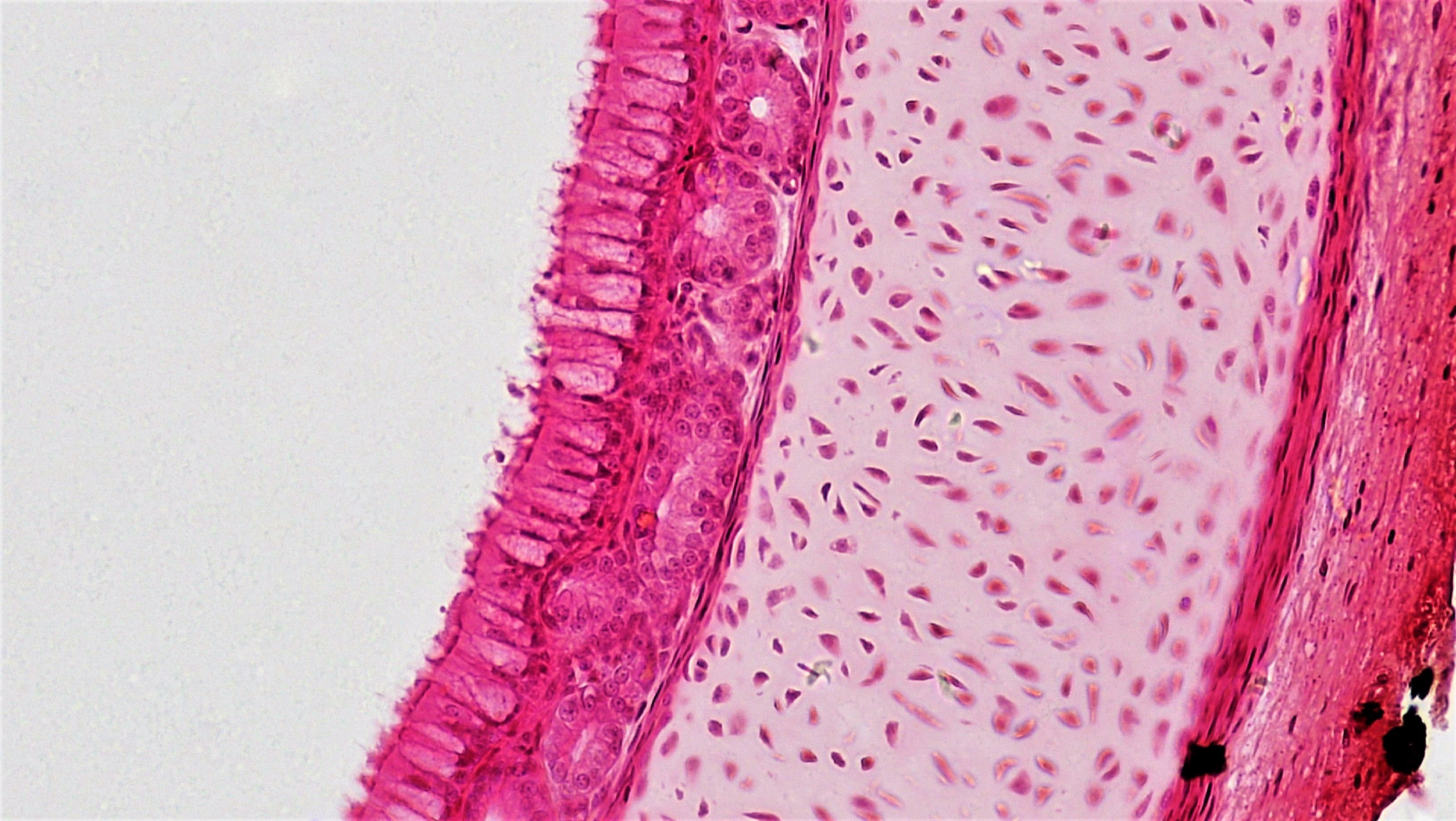
Pseudostratified Ciliated Columnar Epithelium Drawing - The nuclei of these epithelial cells are at different levels leading to the illusion of being stratified. Ciliated epithelia are more common and lines the trachea, bronchi. All of the cells are in contact with the basement membrane (on the. Pseudostratified ciliated columnar epithelium with goblet cells tissue / organ: Web ciliated pseudostratified columnar epithelium. Once again, the bar shows you the thickness of the ciliated pseudostratified epithelium. Web pseudostratified ciliated columnar epithelium. Web pseudostratified columnar epithelia are tissues formed by a single layer of cells that give the appearance of being made from multiple layers, especially when seen in cross section. As implied by their moniker, columnar epithelial cells are taller than they are wide, appearing like numerous miniature pillars. As its name suggests, this epithelium has a false multilayered appearance, when it is in fact a single layer of cells. The cells in this tissue are very tall and thin, and are not all the same shape. Pseudostratified columnar epithelium location or examples. Pseudostratified columnar epithelium location and examples. Web table of contents. Click on links to move to a specific region. Now you can see the individual cells. Web pseudostratified columnar epithelium is found in the respiratory tract, where some of these cells have cilia. 9,523 x 6,085 pixels 216 mb. The two layers of nuclei that will be most apparent are a layer of basal cells, and a layer of columnar cells. 7.3k views 2 years ago cell biology. As implied by their moniker, columnar epithelial cells are taller than they are wide, appearing like numerous miniature pillars. Both simple and pseudostratified columnar epithelia are heterogeneous epithelia because they include additional types of cells interspersed among the epithelial cells. Sorenson university of minnesota minneapolis, mn. Web pseudostratified columnar epithelia are tissues formed by a single layer of cells that. Web ciliated pseudostratified columnar epithelium. If a specimen looks stratified but has cilia, then it is a pseudostratified ciliated epithelium, since stratified epithelia do not have cilia. The image can be changed using any combination of the. Web the epididymus has a pseudostratified columnar epithelium lined by stereocilia. Web pseudostratified columnar epithelia are tissues formed by a single layer of. Now you can see the individual cells. Each cell rests on the basement membrane, although not all cells reach the apical surface. Simple columnar or pseudostratified columnar tissue / organ: Web about press copyright contact us creators advertise developers terms privacy policy & safety how youtube works test new features nfl sunday ticket press copyright. Olfactory epithelium of nasal cavity. 9,523 x 6,085 pixels 216 mb. Functions of the pseudostratified columnar epithelium. Although not always evident, those columnar cells are in contact with the basement membrane. Web 27,160 x 59,070 pixels 6 gb. Web pseudostratified epithelium is a type of simple columnar epithelium. Web pseudostratified columnar epithelia are tissues formed by a single layer of cells that give the appearance of being made from multiple layers, especially when seen in cross section. Once again, the bar shows you the thickness of the ciliated pseudostratified epithelium. Web some cells in the pseudostratified columnar epithelium have cilia on the apical surface that is involved with. Each slide is shown with additional information to its right. Web 27,160 x 59,070 pixels 6 gb. Sorenson university of minnesota minneapolis, mn. Identification of pseudostratified columnar cells under a microscope. The cells in this tissue are very tall and thin, and are not all the same shape. This is the 4th video of our series in which we have. Sorenson university of minnesota minneapolis, mn. The nuclei of these epithelial cells are at different levels leading to the illusion of being stratified. Web pseudostratified columnar epithelia are tissues formed by a single layer of cells that give the appearance of being made from multiple layers, especially when. The image can be changed using any combination of the. Web pseudostratified epithelia function in secretion or absorption. Functions of the pseudostratified columnar epithelium. Now you can see the individual cells. Web pseudostratified columnar epithelia are tissues formed by a single layer of cells that give the appearance of being made from multiple layers, especially when seen in cross section. Web ciliated pseudostratified columnar epithelium. Sorenson university of minnesota minneapolis, mn. Medical school university of minnesota minneapolis, mn. Web pseudostratified columnar epithelium is found in the respiratory tract, where some of these cells have cilia. Web about press copyright contact us creators advertise developers terms privacy policy & safety how youtube works test new features nfl sunday ticket press copyright. Each slide is shown with additional information to its right. As its name suggests, this epithelium has a false multilayered appearance, when it is in fact a single layer of cells. Pseudostratified ciliated columnar epithelium is composed primarily of tall slender cells (columnar cells) with cilia at their apical surface. Based on the presence and absence of cilia, the pseudostratified columnar epithelium has two types: 7.3k views 2 years ago cell biology. Pseudostratified columnar epithelium location or examples. Surface of the pseudostratified columnar epithelium that lines the lumen of the trachea. The cells that comprise the epithelial membranes are variously shaped and are named accordingly. Pseudostratified ciliated columnar epithelium with goblet cells tissue / organ: Ciliated epithelia are more common and lines the trachea, bronchi. The two layers of nuclei that will be most apparent are a layer of basal cells, and a layer of columnar cells.
Pseudostratified Ciliated Columnar / / The cells that comprise the

Pseudostratified columnar epithelium Servier Medical Art

Pseudostratified Columnar Epithelium Diagram

Pseudostratified Ciliated Columnar / / The cells that comprise the

Illustration depicting Pseudostratified Ciliated Columnar Epithelium

Pseudostratified Columnar Epithelium Kit Ng, Ph.D.

How to draw Pseudostratified columnar epithelium most easy way
Pseudostratified Columnar Epithelium Diagram

Pseudostratified Columnar Epithelium Kit Ng, Ph.D.
Pseudostratified columnar ciliated epithelium with goblet cells and
Web Pseudostratified Columnar Epithelium Is Found In The Respiratory Tract, Where Some Of These Cells Have Cilia.
The Cells In This Tissue Are Very Tall And Thin, And Are Not All The Same Shape.
If A Specimen Looks Stratified But Has Cilia, Then It Is A Pseudostratified Ciliated Epithelium, Since Stratified Epithelia Do Not Have Cilia.
Click On Links To Move To A Specific Region.
Related Post: