Prokaryote Drawing
Prokaryote Drawing - Many also have a capsule or slime layer made of polysaccharide. 2.2.2 annotate the diagram from 2.2.1 with the functions of each named structure. Cell components prokaryotic & eukaryotic cells. These cells are structurally simpler and smaller. Web thanks for watching!i am demonstrating the colorful diagram of prokaryotic cells step by step which you can draw very easily. The most common shapes are helices, spheres, and rods (see figure below). It helps in moisture retention, protects the cell when engulfed, and helps in the attachment of cells to nutrients and surfaces. Web do you want to learn how to draw a prokaryotic cell in an easy way? Whereas eukaryotic cells have many different functional compartments, divided by membranes, prokaryotes only. Protects the cell from the outside environment and maintains the shape of the cell.it also prevents the cell from bursting if internal. Nucleoid is spelt wrong in the above video, apologies for any confusion. Although they are tiny, prokaryotic cells can be distinguished by their shapes. A t t students will then be asked to provide a gesture, a sound, or another word to. All prokaryotic cells are encased by a cell wall. Whereas eukaryotic cells have many different functional compartments, divided. These neat, well labelled and. Diagram of a typical prokaryotic cell. The features of a typical prokaryotic cell are shown. Coli) as an example of a prokaryote. Updated on october 30, 2019. Protects the cell from the outside environment and maintains the shape of the cell.it also prevents the cell from bursting if internal. Diagram of a typical prokaryotic cell. These neat, well labelled and. ️ ️ ️ and do tell. Web how to draw a prokaryotic cell for yr 11 ib biologynote: Cells vary regarding other components. Web a prokaryotic cell structure is as follows: Web parts, functions & diagrams of prokaryotes. Protects the cell from the outside environment and maintains the shape of the cell.it also prevents the cell from bursting if internal. It helps in moisture retention, protects the cell when engulfed, and helps in the attachment of cells to. Write in the similarities and differences between prokaryotic and eukaryotic cells. Web prokaryotic cells 2.2.1 draw and label a diagram of the ultrastructure of escherichia coli (e. Web step by step and simple way to draw prokaryotic cell with easy methods.reference; Has has plasma plasma membrane membrane. As i go, i give tips on drawing the various structures. Prokaryotic cell structure and function | channels for pearson+. Vector seamless pattern on the theme of chemistry, biology, genetics, medicine. Typical prokaryotic cells range from 0.1 to 5.0 micrometers (μm) in diameter and are significantly smaller than eukaryotic cells, which usually have diameters ranging from 10 to 100 μm. Nucleoid is spelt wrong in the above video, apologies for any. Coli) as an example of a prokaryote. Web how to draw a prokaryotic cell for yr 11 ib biologynote: Updated on october 30, 2019. Vector seamless pattern on the theme of chemistry, biology, genetics, medicine. Web thanks for watching!i am demonstrating the colorful diagram of prokaryotic cells step by step which you can draw very easily. Although they are tiny, prokaryotic cells can be distinguished by their shapes. The features of a typical prokaryotic cell are shown. Most prokaryotic cells are much smaller than eukaryotic cells. It helps in moisture retention, protects the cell when engulfed, and helps in the attachment of cells to nutrients and surfaces. All prokaryotic cells are encased by a cell wall. Cell components prokaryotic & eukaryotic cells. Whereas eukaryotic cells have many different functional compartments, divided by membranes, prokaryotes only. Prokaryotic cell structure and function | channels for pearson+. As i go, i give tips on drawing the various structures. Diagram of a typical prokaryotic cell. Web step by step and simple way to draw prokaryotic cell with easy methods.reference; Web prokaryotic cell diagram and facts. These cells are structurally simpler and smaller. Web parts, functions & diagrams of prokaryotes. The most common shapes are helices, spheres, and rods (see figure below). General science, 7th standard text book.this video explains how to draw pro. [2] the word prokaryote comes from the ancient greek πρό ( pró) 'before' and κάρυον ( káruon) 'nut, kernel'. The figure below shows the sizes of prokaryotic, bacterial, and eukaryotic, plant and animal, cells as well as other molecules and organisms on a. Web step by step and simple way to draw prokaryotic cell with easy methods.reference; ️ ️ ️ and do tell. It helps in moisture retention, protects the cell when engulfed, and helps in the attachment of cells to nutrients and surfaces. Prokaryotic cells are much simpler than the more evolutionarily advanced. Web prokaryotic cells 2.2.1 draw and label a diagram of the ultrastructure of escherichia coli (e. Recall that prokaryotes are divided into two different domains, bacteria and archaea, which together with eukarya, comprise the three domains of life (figure 27.2.3 27.2. The three most common prokaryotic cell shapes are shown here. These cells are structurally simpler and smaller. Web parts, functions & diagrams of prokaryotes. Bacteria and archaea are both prokaryotes but differ enough to be. Most prokaryotic cells are much smaller than eukaryotic cells. Prokaryotic cells are much smaller than eukaryotic cells, have no nucleus, and lack organelles. Web i draw a bacterial cell to show you how to make an accurate biological drawing of a prokaryotic cell.
Prokaryotic Cell Structure Corresponding Designations Microbiology
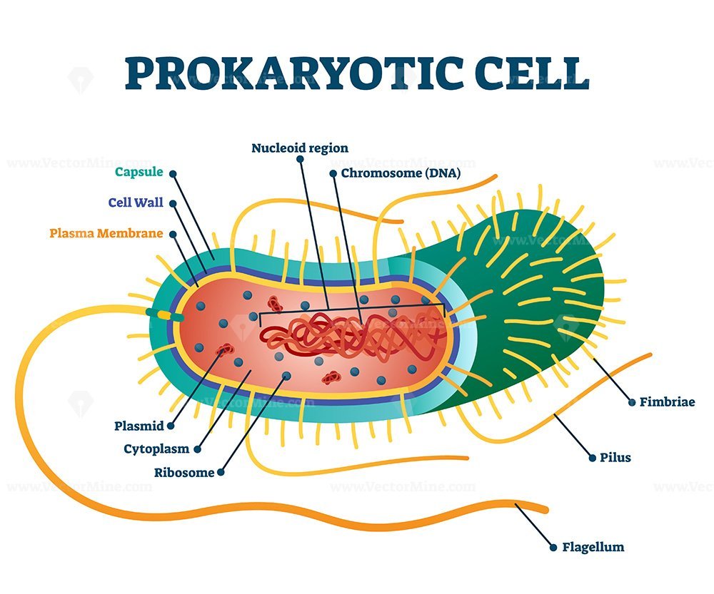
Prokaryotic Cells Labeled

How to draw a prokaryotic cell prokaryotic organism Bacterial cell

Prokaryote Wikipedia

Prokaryotes Intro at Austin Community College StudyBlue
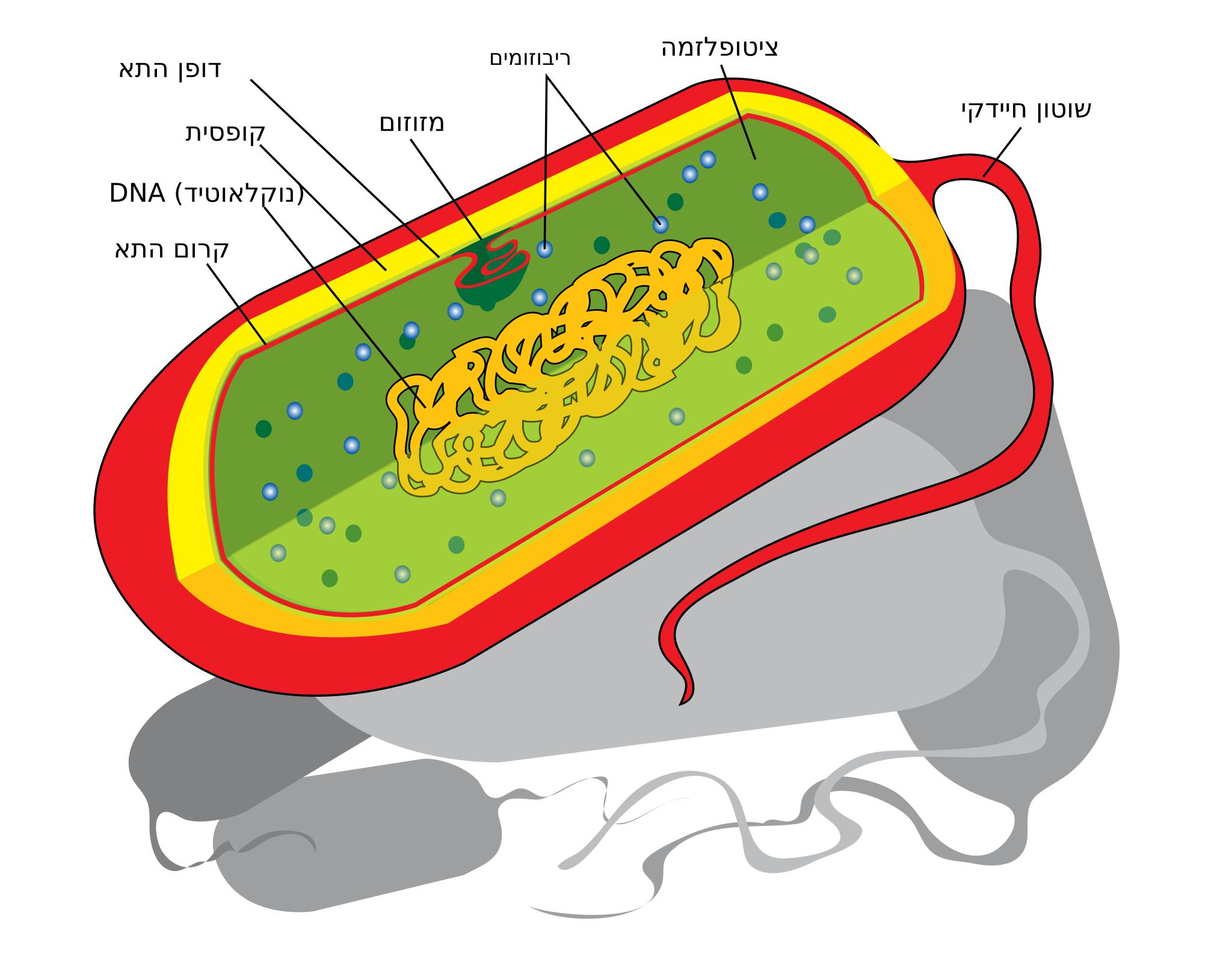
Free Images prokaryote cell diagram he
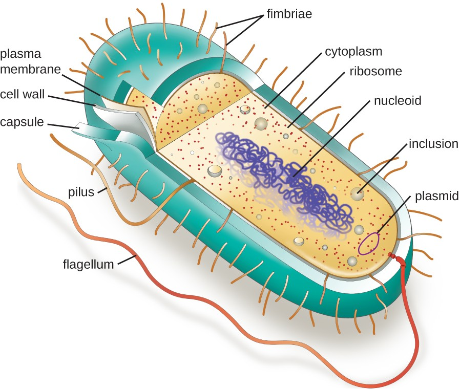
2.3 Unique Characteristics of Prokaryotic Cells Allied Health
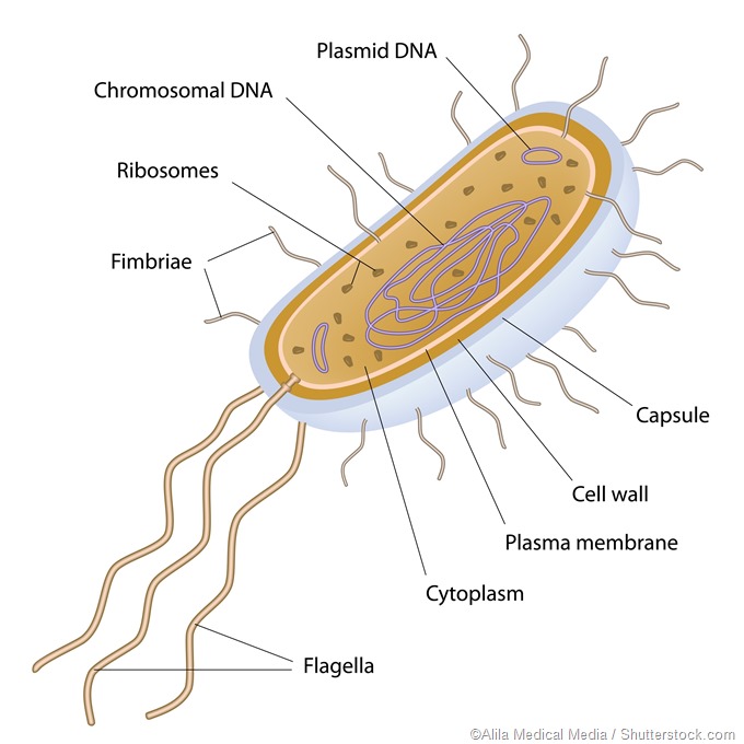
Prokaryotes
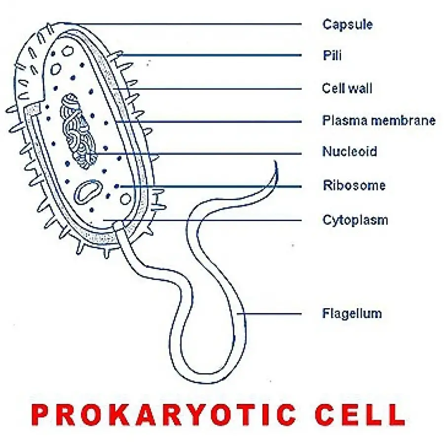
Simple Prokaryotic Cell Diagram
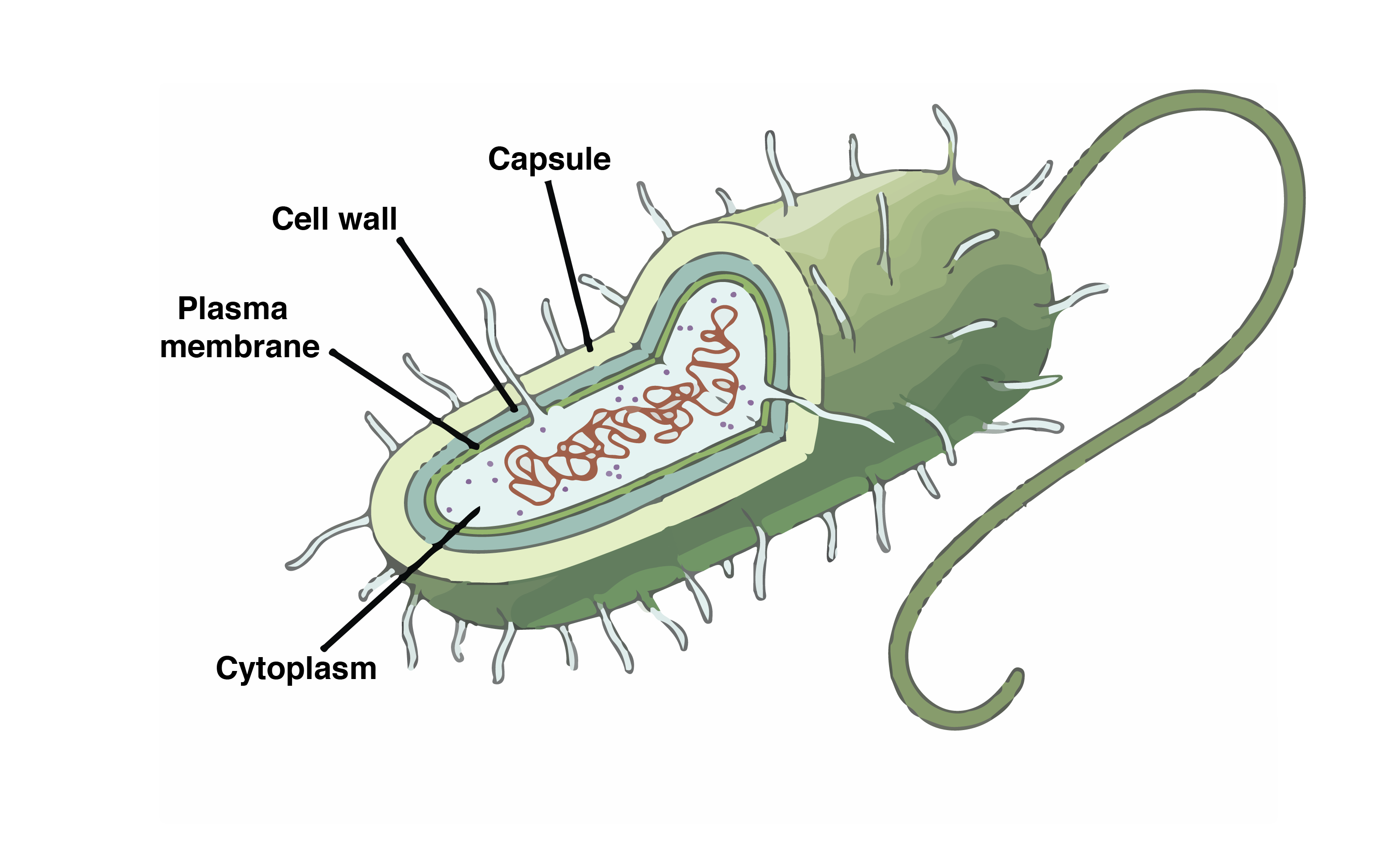
Learning Through Art Structures of a Prokaryotic Cell
Eukaryotic Cells Are Generally Much Larger, Between 10 And 100 Micrometers.
Prokaryotic Cell Structure And Function | Channels For Pearson+.
Cell Components Prokaryotic & Eukaryotic Cells.
2.2.2 Annotate The Diagram From 2.2.1 With The Functions Of Each Named Structure.
Related Post: