Pediatric X Ray Technique Chart
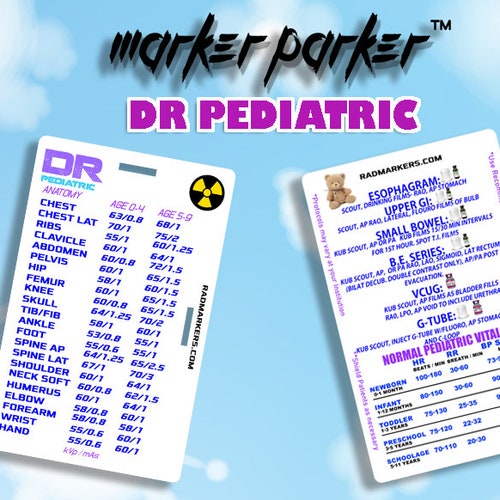
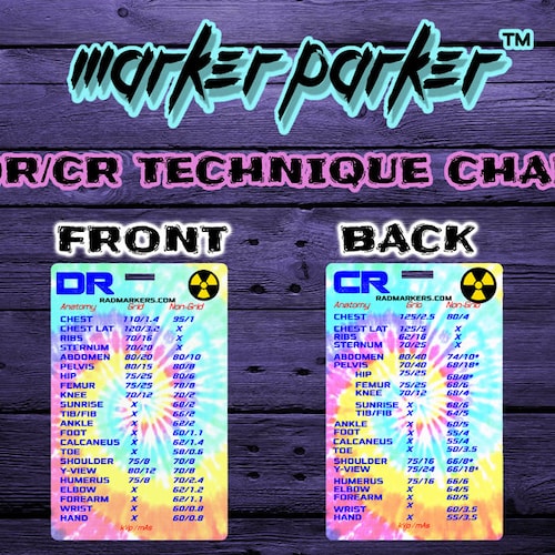
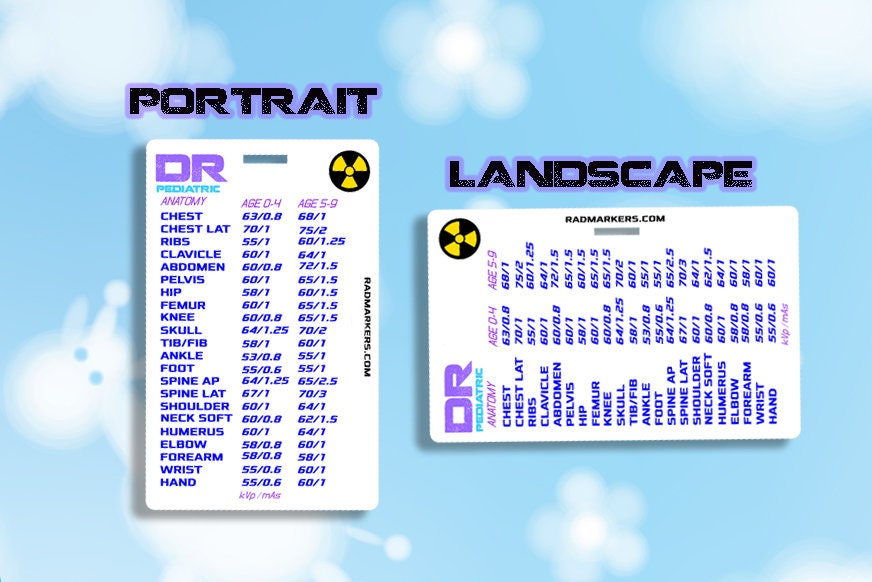
Pediatric X Ray Technique Chart - Patient is placed on top of the detector. As radiation protection is necessary for pediatric patients, it is essential to image the chest properly and avoid unnecessary repeats. Entire lung fields should be visible; Skeletal system of the chest 9. Used medical accessories (tube, drains, catheters etc) normal chest x. Exposure, imaging, paediatric, radiography, technique. This study aimed to identify proper exposure techniques to maintain optimal diagnostic image quality with minimum radiation dose. What is the importance of radiographic image quality? It also works for a suitable portable x ray techniques chart. The ap erect view is often chosen over the pa erect view for younger children as this view allows for observing the child’s breathing and decreased patient stress (due to the child being able to observe what is happening in the room). Department of medical imaging, royal children’s hospital, brisbane, queensland, australia. Patient is placed on top of the detector. Dl, yates ar, zuppa af, pollack mm (2020). Web it is best practice, when imaging a pediatric patient, to use exposure techniques that are appropriate for the size of the patient and to properly position and immobilize the patient to avoid repeat. Confirming the location of line placement (e.g. Web performing chest radiography on pediatric patients can be for a number of indications 1: Pediatric radiography is a subset of general radiography specializing in the radiographic imaging of the pediatric population. Respiratory distress syndrome) cardiac disease. Web 24 cm x 30 cm or 35 cm x 43 cm depending on the patient’s. Dl, yates ar, zuppa af, pollack mm (2020). Inferior to the inferior pubic rami The general principles of radiography remain the same. Web this view is preferred in infant and neonate imaging, whilst ap erect and pa erect views are ideal for children able to cooperate in sitting or standing 1. Respiratory distress syndrome) cardiac disease. Dl, yates ar, zuppa af, pollack mm (2020). Inferior to the inferior pubic rami Skeletal system of the chest 9. An image produced resulting in nondiagnostic quality is useless to. This study aimed to identify proper exposure techniques to maintain optimal diagnostic image quality with minimum radiation dose. This study aimed to identify proper exposure techniques to maintain optimal diagnostic image quality with minimum radiation dose. This card has dr techniques on the front and fluoroscopy techniques and vitals on the back of the card. Entire lung fields should be visible; Web last revised by andrew murphy on 23 mar 2023. An image produced resulting in nondiagnostic quality. Web last revised by andrew murphy on 23 mar 2023. The midsagittal plane (xiphisternum) at the level of the iliac crest. Exposure, imaging, paediatric, radiography, technique. However, the ap view will result in an increased radiation dose to radiosensitive organs. Heart and lower mediastinum 7. This study aimed to identify proper exposure techniques to maintain optimal diagnostic image quality with minimum radiation dose. Laterally to the lateral abdominal wall. Skeletal system of the chest 9. Heart and lower mediastinum 7. Exposure, imaging, paediatric, radiography, technique. Dl, yates ar, zuppa af, pollack mm (2020). Pediactric technique chart with pediatric fluoroscopy protocols and baseline vital chart. What is the importance of radiographic image quality? Inferior to the inferior pubic rami This study aimed to identify proper exposure techniques to maintain optimal diagnostic image quality with minimum radiation dose. You attach the card to a badge reel or your existing badge to have available at your fingertips should you need help selecting a. An image produced resulting in nondiagnostic quality is useless to. Confirming the location of line placement (e.g. Respiratory distress syndrome) cardiac disease. However, the ap view will result in an increased radiation dose to radiosensitive organs. This card has dr techniques on the front and fluoroscopy techniques and vitals on the back of the card. This study aimed to identify proper exposure techniques to maintain optimal diagnostic image quality with minimum radiation dose. A dentist or hygienist should first examine children’s teeth before deciding on the number and types of radiographs. You attach the card to. Confirming the location of line placement (e.g. Department of medical imaging, royal children’s hospital, brisbane, queensland, australia. This card has dr techniques on the front and fluoroscopy techniques and vitals on the back of the card. It also works for a suitable portable x ray techniques chart. Web 24 cm x 30 cm or 35 cm x 43 cm depending on the patient’s size. An image produced resulting in nondiagnostic quality is useless to. Web this view is preferred in infant and neonate imaging, whilst ap erect and pa erect views are ideal for children able to cooperate in sitting or standing 1. Laterally to the lateral abdominal wall. Inferior to the inferior pubic rami Skeletal system of the chest 9. What is the importance of radiographic image quality? Pediactric technique chart with pediatric fluoroscopy protocols and baseline vital chart. The general principles of radiography remain the same. You attach the card to a badge reel or your existing badge to have available at your fingertips should you need help selecting a. Entire lung fields should be visible; This study aimed to identify proper exposure techniques to maintain optimal diagnostic image quality with minimum radiation dose.
Pediatric Chest X Ray Technique Chart

Pediatric Chest X Ray Technique Chart

Pediatric X Ray Technique Chart

Pediatric X Ray Technique Chart

Pediatric Chest X Ray Technique Chart Labb by AG

Technique Charts for Radiologic Technologists Exposure Guides

PEDIATRIC DR Technique Chart for Xray Technologist Etsy

A paediatric Xray exposure chart Semantic Scholar

A paediatric Xray exposure chart Semantic Scholar

Pediatric Chest X Ray Technique Chart Labb by AG
Web It Is Best Practice, When Imaging A Pediatric Patient, To Use Exposure Techniques That Are Appropriate For The Size Of The Patient And To Properly Position And Immobilize The Patient To Avoid Repeat Exposures.
However, The Ap View Will Result In An Increased Radiation Dose To Radiosensitive Organs.
Patient Is Placed On Top Of The Detector.
The Number And Types Of Radiographs Necessary
Related Post: