Myotomal Pattern
Myotomal Pattern - This term is based on the combination of two ancient greek roots; A myotome is a group of muscles innervated by the ventral root a single spinal nerve. Motor loss may occur in a myotomal pattern (table (table1). From day 20 onwards the paraxial mesoderm begins to. Every fiber that is part of a motor unit contracts (shortens) to move when its respective nerve is. [2] in vertebrate embryonic development, a myotome is the part of a somite that develops into muscle. The area directly adjacent to the neural tube is known as the paraxial mesoderm. A myotome is the group of muscles that a single spinal nerve innervates. The distribution of pain and motor findings on physical exam should guide the neurosurgeon to the region of the spine to focus on,. Each nerve cell innervates (provides signals to) several muscle fibers. Myotome testing is an essential part of neurological examination when suspecting radiculopathy. Myotomes are separated by myosepta (singular: Each nerve cell innervates (provides signals to) several muscle fibers. Skeletal muscle development can be traced to the appearance of somites.by day 20 the trilaminar disc has formed and the mesoderm has differentiated into different areas. The inguinal region and the very. The distribution of pain and motor findings on physical exam should guide the neurosurgeon to the region of the spine to focus on,. Every fiber that is part of a motor unit contracts (shortens) to move when its respective nerve is. Each nerve cell innervates (provides signals to) several muscle fibers. Web anatomy and function of the peripheral nervous system.. Web anatomy and function of the peripheral nervous system. Web dermatome map of the torso dermatomes of the lower limb. Web classically, when radiculopathy is caused by nerve root compression pain, sensory loss occurs in a dermatomal pattern (figure (figure1). Motor loss may occur in a myotomal pattern (table (table1). Myotomes are much more complex to test then dermatomes, since. Web origin of myotomes. Each nerve cell innervates (provides signals to) several muscle fibers. This term is based on the combination of two ancient greek roots; Skeletal muscle development can be traced to the appearance of somites.by day 20 the trilaminar disc has formed and the mesoderm has differentiated into different areas. Web classically, when radiculopathy is caused by nerve. A myotome is a group of muscles innervated by the ventral root a single spinal nerve. The area directly adjacent to the neural tube is known as the paraxial mesoderm. The lateral aspect of the calcaneus. Skeletal muscle development can be traced to the appearance of somites.by day 20 the trilaminar disc has formed and the mesoderm has differentiated into. The middle and lateral aspect of the anterior thigh. Every fiber that is part of a motor unit contracts (shortens) to move when its respective nerve is. Web dermatome map of the torso dermatomes of the lower limb. Myo=muscle, tome = a section, volume) is defined as a group of muscles which is innervated by single spinal nerve root. Web. From day 20 onwards the paraxial mesoderm begins to. The area directly adjacent to the neural tube is known as the paraxial mesoderm. The distribution of pain and motor findings on physical exam should guide the neurosurgeon to the region of the spine to focus on,. Myotomes are separated by myosepta (singular: Web this video is a great of learning. The dorsum of the foot at the third metatarsophalangeal joint. Myotomes are much more complex to test then dermatomes, since each skeletal muscle is innervated by nerves. A myotome is the group of muscles that a single spinal nerve innervates. A myotome is a group of muscles innervated by the ventral root a single spinal nerve. Web classically, when radiculopathy. Skeletal muscle development can be traced to the appearance of somites.by day 20 the trilaminar disc has formed and the mesoderm has differentiated into different areas. Each nerve cell innervates (provides signals to) several muscle fibers. Web origin of myotomes. From day 20 onwards the paraxial mesoderm begins to. The distribution of pain and motor findings on physical exam should. Skeletal muscle development can be traced to the appearance of somites.by day 20 the trilaminar disc has formed and the mesoderm has differentiated into different areas. Myotome testing is an essential part of neurological examination when suspecting radiculopathy. The lateral aspect of the calcaneus. This term is based on the combination of two ancient greek roots; Motor loss may occur. The lateral aspect of the calcaneus. Myo=muscle, tome = a section, volume) is defined as a group of muscles which is innervated by single spinal nerve root. Web we have described split finger, that is, weaker fdp1 than fdp4, as a characteristic feature of als.59 the pattern is opposite between distal csa and als, and may be useful in differentiating the two disorders. Myotome testing is an essential part of neurological examination when suspecting radiculopathy. A myotome is a group of muscles innervated by the ventral root a single spinal nerve. Web origin of myotomes. Myotomes are separated by myosepta (singular: The inguinal region and the very top of the medial thigh. The dorsum of the foot at the third metatarsophalangeal joint. A single nerve and its corresponding muscle fibers comprise a motor unit. From day 20 onwards the paraxial mesoderm begins to. The area directly adjacent to the neural tube is known as the paraxial mesoderm. Like spinal nerves, myotomes are organised into. The distribution of pain and motor findings on physical exam should guide the neurosurgeon to the region of the spine to focus on,. Motor loss may occur in a myotomal pattern (table (table1). A myotome is the group of muscles that a single spinal nerve innervates.
Dermatomes, Myotomes and DTR Nervous System Poster 18 X 24 Chiropractic

Dermatomes And Myotomes Chart And Map

Myotomes And Dermatomes Map

How To Learn Dermatomes Permissiondeath

Dermatomes and myotomes Dermatomes and myotomes, Physical therapy
![Dermatomes and Myotomes [The Comprehensive Guide 2023] Physio Health](https://physiohealthexpert.com/wp-content/uploads/2023/01/Dermatomes-and-Myotomes.jpg)
Dermatomes and Myotomes [The Comprehensive Guide 2023] Physio Health

Dermatomes and Myotomes Jonathan Collier Injury Rehabilitation

26e81966628d70fe0f1e6a21f7c9bff6bb50d626 742×559 pixels Physical

Myotome map to remember muscle roots + dermatome signature zones medicine

Lower Extremity Dermatomes and Myotomes Reflexes GrepMed
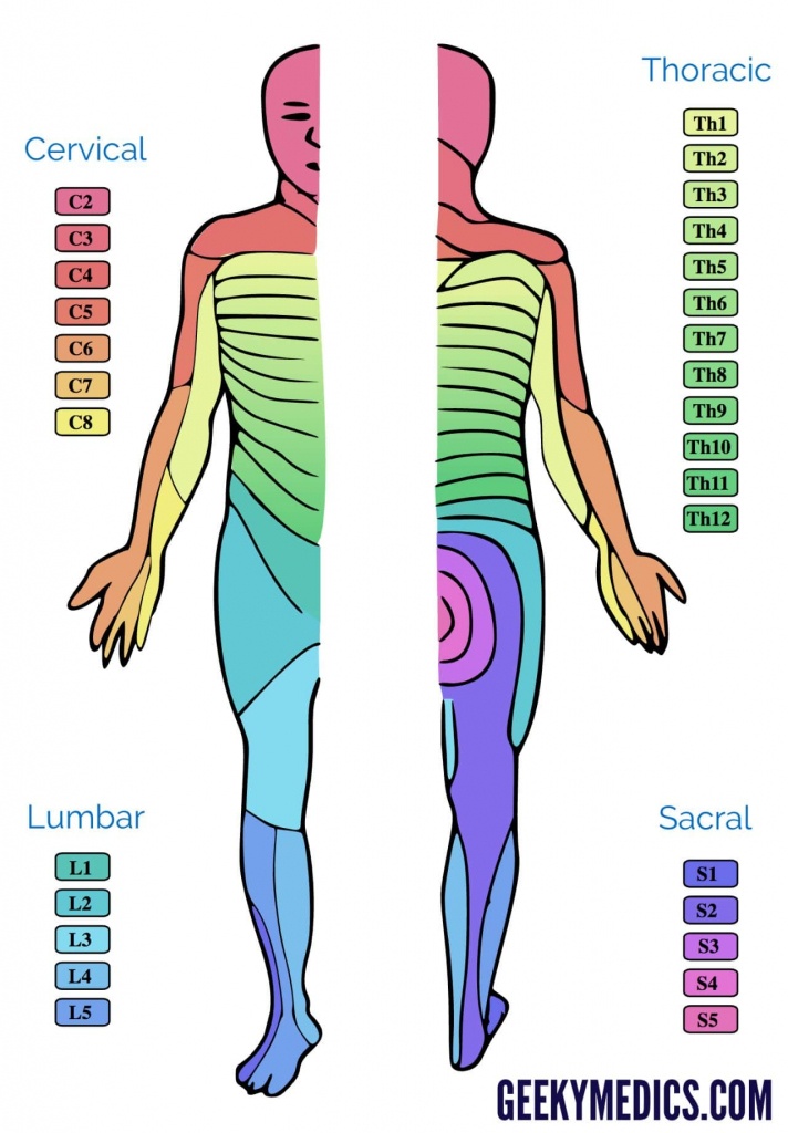
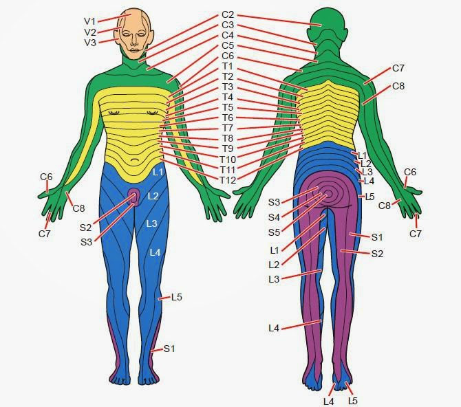
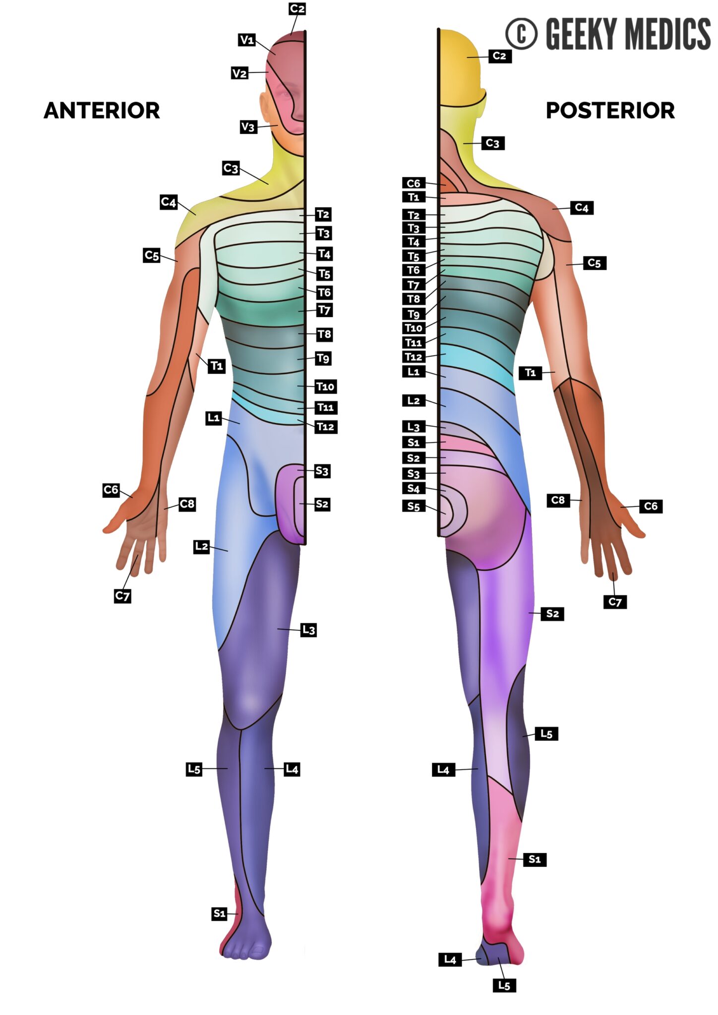
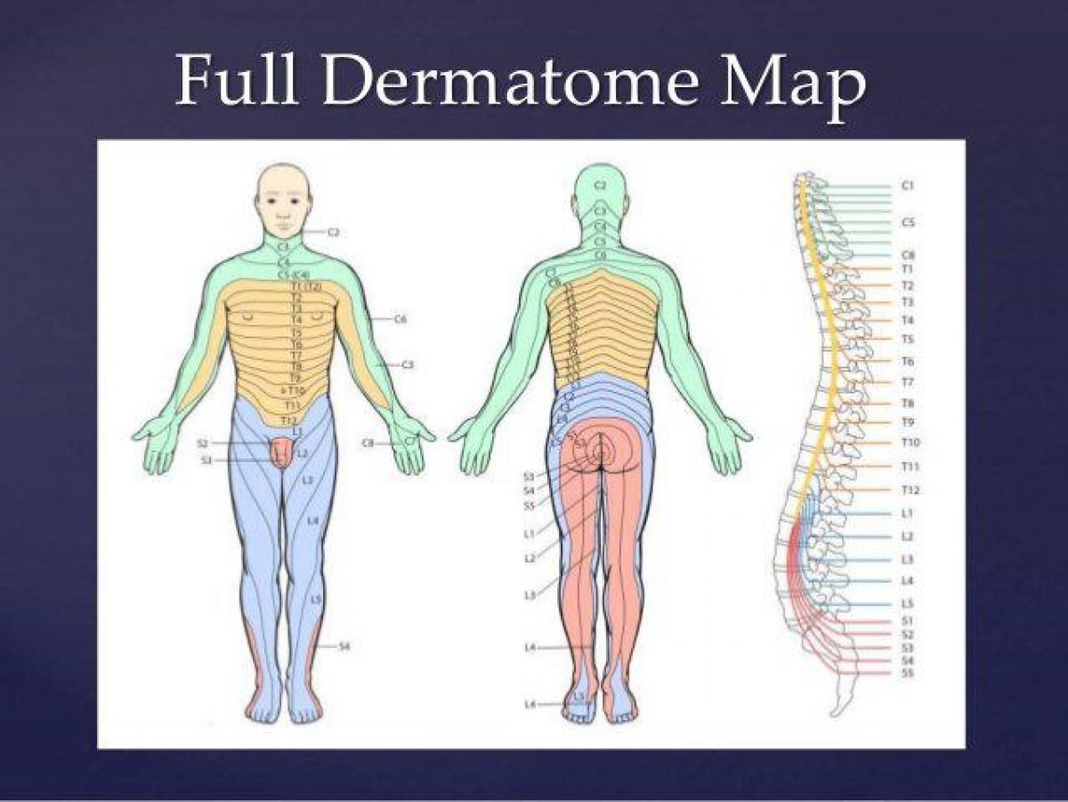
Web Dermatome Map Of The Torso Dermatomes Of The Lower Limb.
The Medial Epicondyle Of The Femur.
Web Classically, When Radiculopathy Is Caused By Nerve Root Compression Pain, Sensory Loss Occurs In A Dermatomal Pattern (Figure (Figure1).
Web This Video Is A Great Of Learning The Myotomes Of The Upper Limb, And Crucially, How To Test Them In Clinical Practice!⭐ Membership:
Related Post: