Membrane Drawing
Membrane Drawing - Web the plasma membrane of a cell is a network of lipids and proteins that forms the boundary between a cell’s contents and the outside of the cell. Web the cell membrane, also called the plasma membrane, is a thin layer that surrounds the cytoplasm of all prokaryotic and eukaryotic cells, including plant and animal cells. Regarding osmotic membrane bioreactor (ombr), special attention is needed for the selection of a proper draw solution, which desirably should be low cost, have high osmolality, reduce reverse salt flux, and can be easily reconcentrated. Of course, a cell is ever so much more than just a bag of goo. Davson and danielli theorized that the plasma membrane’s structure. The phospholipids are tightly packed together, and the membrane has a hydrophobic interior. The head is a phosphate molecule that is. This was the first model that others in the scientific community widely accepted. The head and the two tails. It was based on the plasma membrane’s “railroad track” appearance in early electron micrographs. It's a complex, highly organized unit, the basic building block of all living things. Web in most resting neurons, the potential difference across the membrane is about 30 to 90 mv (a mv is 1 / 1000 of a volt), with the inside of the cell more negative than the outside. This was the first model that others in the. Web the plasma membrane of a cell is a network of lipids and proteins that forms the boundary between a cell’s contents and the outside of the cell. This was the first model that others in the scientific community widely accepted. Use the lace cording chromosomes to model the g2 phase of interphase (after each chromosome was replicated during s. As a comparison, human red blood cells, visible via light microscopy, are approximately 8 μm thick, or approximately 1,000 times thicker than a plasma membrane. The plasma membrane protects intracellular components from the extracellular environment. Web when drawing and labeling a diagram of the plasma membrane you should be sure to include:the phospholipid bilayer with hydrophobic 'tails' and hydrophilic 'h.. Web on the paper draw the cell membrane, nucleus, nucleolus, centrioles. Doing this will help you to understand where things are in a cell and why they are in specific positions. Thus preventing them from drawing positive ions. The plasma membrane protects intracellular components from the extracellular environment. Cells are the basis unit of life. Chemical and biomedical engineers study cells and their composition as part of. Web membranes 2.4.1 draw and label a diagram to show the structure of membranes. Specialized structure that surrounds the cell and its internal environment; The absence of ions in the secreted mucus. Thus preventing them from drawing positive ions. Web in 1935, hugh davson and james danielli proposed the plasma membrane’s structure. Specialized structure that surrounds the cell and its internal environment; It's a complex, highly organized unit, the basic building block of all living things. The fluid mosaic model of the plasma membrane structure describes the plasma membrane as a fluid combination of. That is, neurons have a. Web for students doing ib biology. It separates the cytoplasm (the contents of the cell) from the external environment. 2.4.2 explain how the hydrophobic and hydrophilic properties of phospholipids help to maintain the structure of cell membranes. Molecule that contains both a hydrophobic and a hydrophilic end. It's a complex, highly organized unit, the basic building block of all living. There are two important parts of a phospholipid: Web in 1935, hugh davson and james danielli proposed the plasma membrane’s structure. Cells are the basis unit of life. The main function of the plasma membrane is to protect the cell from its surrounding environment. Phospholipid molecules make up the cell membrane and are hydrophilic (attracted to. Cells exclude some substances, take in others, and excrete still others, all in controlled quantities. Of course, a cell is ever so much more than just a bag of goo. It was based on the plasma membrane’s “railroad track” appearance in early electron micrographs. Molecule that contains both a hydrophobic and a hydrophilic end. Use white beads to represent centromeres. And the plasma membrane and cytoplasm are actually pretty sophisticated. Thus preventing them from drawing positive ions. Follow along and draw the plasma membrane.music: The head is a phosphate molecule that is. The head and the two tails. Its function is to protect the integrity of the interior of the cell by allowing certain substances into the cell while keeping other substances out. Web the cell membrane is made up of phospholipids, which have a hydrophilic phosphate head, a glycerol backbone, and two hydrophobic fatty acid tails. The principal components of the plasma membrane are lipids ( phospholipids and cholesterol), proteins, and carbohydrates. Specialized structure that surrounds the cell and its internal environment; It also serves as a base of attachment for the cytoskeleton in some. Of course, a cell is ever so much more than just a bag of goo. It was based on the plasma membrane’s “railroad track” appearance in early electron micrographs. A 3d diagram of the cell membrane. The absence of ions in the secreted mucus. Plasma membranes enclose the borders of cells, but rather than being a static bag, they are dynamic and constantly in flux. This was the first model that others in the scientific community widely accepted. And the plasma membrane and cytoplasm are actually pretty sophisticated. The head and the two tails. Molecule that contains both a hydrophobic and a hydrophilic end. Doing this will help you to understand where things are in a cell and why they are in specific positions. Phospholipid molecules make up the cell membrane and are hydrophilic (attracted to.
How to Draw Cell Membrane Fluid Mosaic Model Diagram Step by Step
:max_bytes(150000):strip_icc()/cell-membrane-373364_final-5b5f300546e0fb008271ce52.png)
Cell Membrane Function and Structure
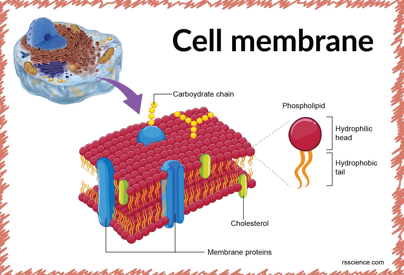
Cell membrane definition, structure, function, and biology
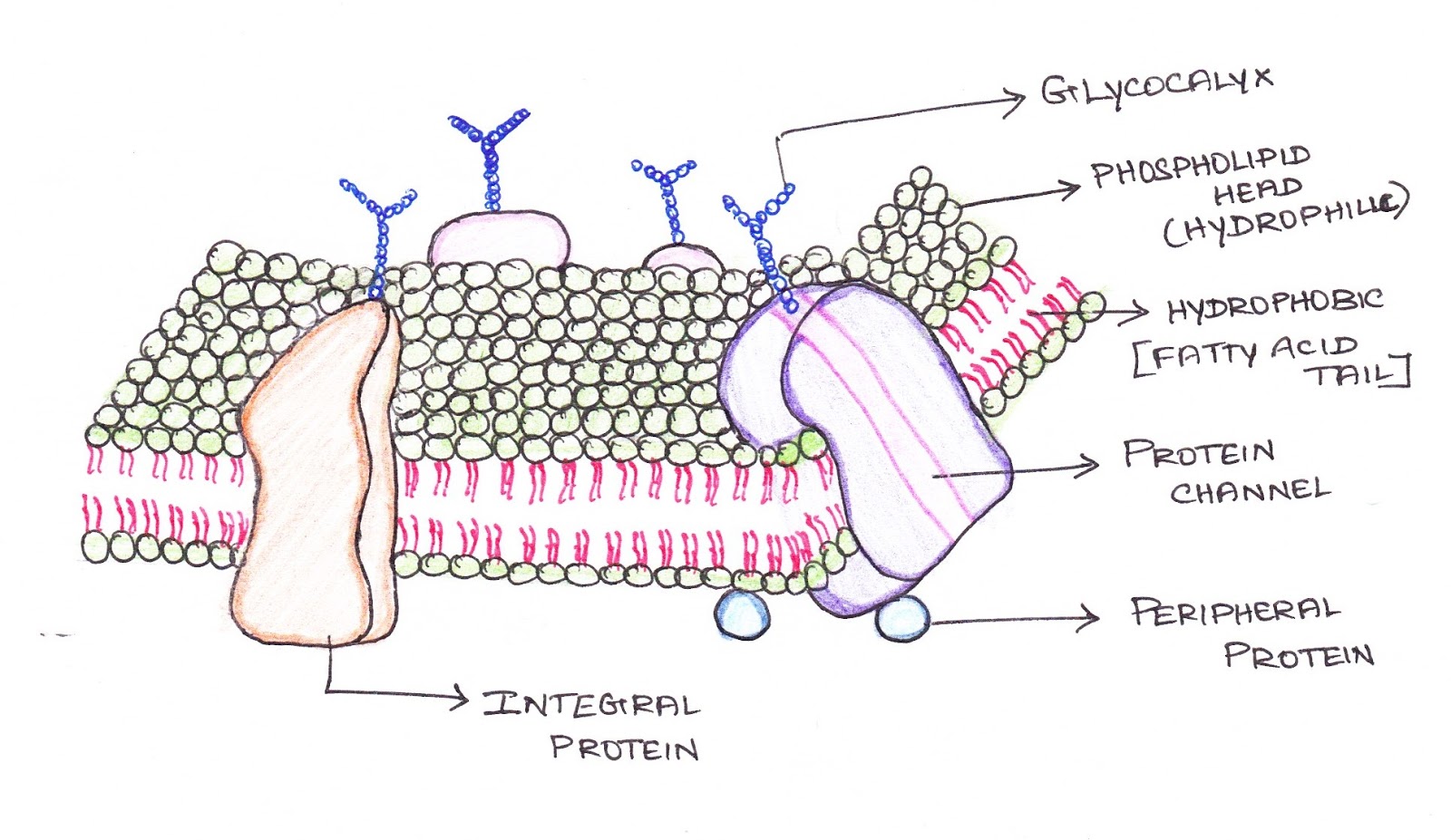
Cell Membrane Drawing Project at GetDrawings Free download

TJ. Schematic diagram of typical membrane proteins in a biological

DRAW IT NEAT How to draw plasma membrane (Cell membrane)
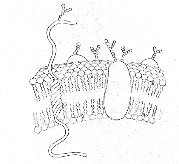
DRAW IT NEAT How to draw plasma membrane (Cell membrane)

Cell membrane with labeled educational structure scheme vector
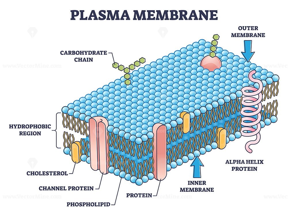
Cell membrane or cytoplasmic membrane microscopic structure outline
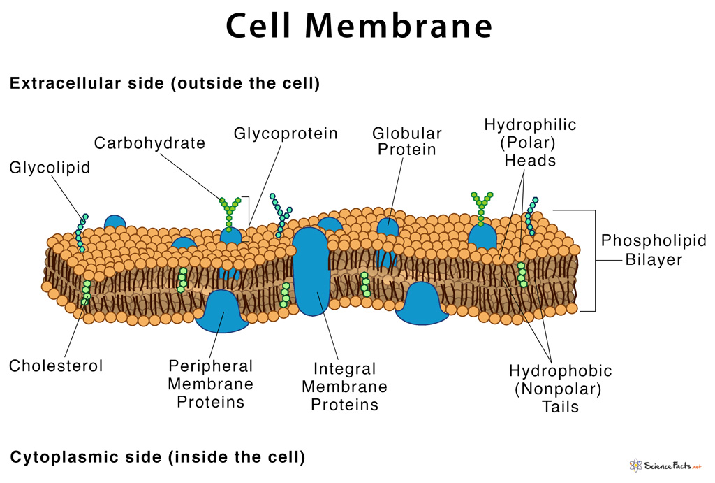
Cell Membrane Definition, Structure, & Functions with Diagram
Web On The Paper Draw The Cell Membrane, Nucleus, Nucleolus, Centrioles.
So As We Mentioned, The Cell Membrane Is Actually A Sphere That Surrounds Our Cell.
The Structure Of The Lipid Bilayer Allows Small, Uncharged Substances Such As Oxygen And Carbon Dioxide, And Hydrophobic Molecules Such As Lipids, To Pass Through The Cell Membrane,.
Web A Cell’s Plasma Membrane Defines The Boundary Of The Cell And Determines The Nature Of Its Contact With The Environment.
Related Post: