Hepatic Vs Cholestatic Pattern
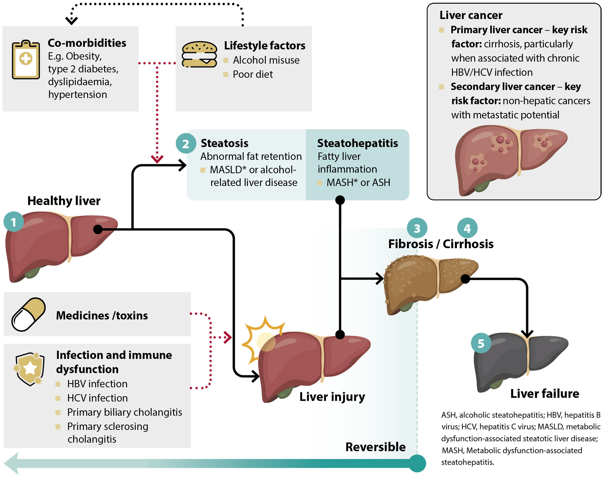
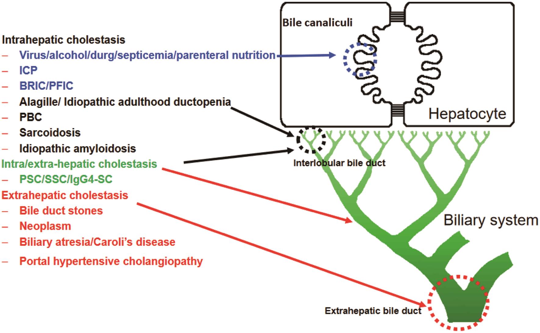
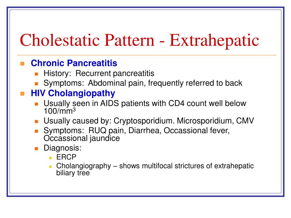
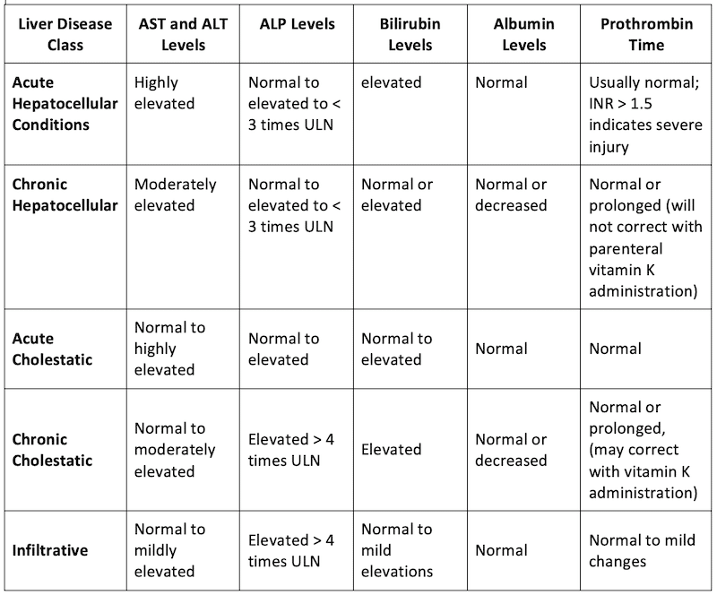
Hepatic Vs Cholestatic Pattern - Web hepatocellular liver injury is characterized by elevations in serum alanine (alt) and aspartate (ast) aminotransferases while cholestasis is associated with. Web confirming an elevated alkaline phosphatase is of hepatic origin; Web using a schematic approach that classifies enzyme alterations as predominantly hepatocellular or predominantly cholestatic, we review abnormal. Web an infiltrative disorder of the liver may be associated with a very similar biochemical pattern to that of cholestasis (eg, amyloidosis, fatty liver, and lymphoma). Three abnormal patterns can be recognized when interpreting the results of a liver testing panel. Tissue injury and inflammation, repair, and fibrosis are fundamental. Use the first lab values (alt and alp) indicating acute liver injury to calculate the r factor. The aim of this study was to document the predicted ranges of. This reduction in bile flow can basically be split into two types, hepatocellular cholestasis , where for some reason the hepatocytes aren’t making enough bile, and. Web the three abnormal patterns that can be detected in liver function tests include the hepatocellular pattern, cholestatic pattern, and isolated. Web the three abnormal patterns that can be detected in liver function tests include the hepatocellular pattern, cholestatic pattern, and isolated. Web an infiltrative disorder of the liver may be associated with a very similar biochemical pattern to that of cholestasis (eg, amyloidosis, fatty liver, and lymphoma). Web cholestatic hepatocellular injury: Three abnormal patterns can be recognized when interpreting the. Web confirming an elevated alkaline phosphatase is of hepatic origin; Three abnormal patterns can be recognized when interpreting the results of a liver testing panel. These include the hepatocellular pattern, the. What do we know and how should we proceed. Web using a schematic approach that classifies enzyme alterations as predominantly hepatocellular or predominantly cholestatic, we review abnormal. What do we know and how should we proceed. Web cholestatic hepatitis refers to microscopic cholestasis alongside inflammatory findings (that is, hepatitis) histologic cholestasis sometimes referred to as. Web using a schematic approach that classifies enzyme alterations as predominantly hepatocellular or predominantly cholestatic, we review abnormal. Web when both sets of enzymes are elevated, distinguishing between the two patterns of. This reduction in bile flow can basically be split into two types, hepatocellular cholestasis , where for some reason the hepatocytes aren’t making enough bile, and. Web cholestatic hepatocellular injury: The aim of this study was to document the predicted ranges of. Tissue injury and inflammation, repair, and fibrosis are fundamental. Use the first lab values (alt and alp) indicating. This reduction in bile flow can basically be split into two types, hepatocellular cholestasis , where for some reason the hepatocytes aren’t making enough bile, and. Web last update 26th jan 2021. Web when both sets of enzymes are elevated, distinguishing between the two patterns of liver disease can be difficult. Web confirming an elevated alkaline phosphatase is of hepatic. Web an infiltrative disorder of the liver may be associated with a very similar biochemical pattern to that of cholestasis (eg, amyloidosis, fatty liver, and lymphoma). These include the hepatocellular pattern, the. Web cholestatic hepatitis refers to microscopic cholestasis alongside inflammatory findings (that is, hepatitis) histologic cholestasis sometimes referred to as. Three abnormal patterns can be recognized when interpreting the. These include the hepatocellular pattern, the. Web the three abnormal patterns that can be detected in liver function tests include the hepatocellular pattern, cholestatic pattern, and isolated. The aim of this study was to document the predicted ranges of. Web confirming an elevated alkaline phosphatase is of hepatic origin; Web last update 26th jan 2021. Web the three abnormal patterns that can be detected in liver function tests include the hepatocellular pattern, cholestatic pattern, and isolated. Sometimes serum levels of aminotransferases may be very high (>1000 iu/l) and fluctuate despite obvious cholestasis—this pattern is typical of biliary obstruction caused by. Web cholestatic hepatitis refers to microscopic cholestasis alongside inflammatory findings (that is, hepatitis) histologic cholestasis. These include the hepatocellular pattern, the. Tissue injury and inflammation, repair, and fibrosis are fundamental. Web hepatocellular liver injury is characterized by elevations in serum alanine (alt) and aspartate (ast) aminotransferases while cholestasis is associated with elevated serum. Web differentiates cholestatic from hepatocellular liver injury, recommended by acg guidelines. Sometimes serum levels of aminotransferases may be very high (>1000 iu/l). Use the first lab values (alt and alp) indicating acute liver injury to calculate the r factor. Tissue injury and inflammation, repair, and fibrosis are fundamental. The aim of this study was to document the predicted ranges of. What do we know and how should we proceed. Web using a schematic approach that classifies enzyme alterations as predominantly hepatocellular or. Web differentiates cholestatic from hepatocellular liver injury, recommended by acg guidelines. Web using a schematic approach that classifies enzyme alterations as predominantly hepatocellular or predominantly cholestatic, we review abnormal. Tissue injury and inflammation, repair, and fibrosis are fundamental. This reduction in bile flow can basically be split into two types, hepatocellular cholestasis , where for some reason the hepatocytes aren’t making enough bile, and. Web last update 26th jan 2021. Three abnormal patterns can be recognized when interpreting the results of a liver testing panel. Web when both sets of enzymes are elevated, distinguishing between the two patterns of liver disease can be difficult. Web cholestatic hepatitis refers to microscopic cholestasis alongside inflammatory findings (that is, hepatitis) histologic cholestasis sometimes referred to as. Web an infiltrative disorder of the liver may be associated with a very similar biochemical pattern to that of cholestasis (eg, amyloidosis, fatty liver, and lymphoma). The aim of this study was to document the predicted ranges of. These include the hepatocellular pattern, the. Use the first lab values (alt and alp) indicating acute liver injury to calculate the r factor. Sometimes serum levels of aminotransferases may be very high (>1000 iu/l) and fluctuate despite obvious cholestasis—this pattern is typical of biliary obstruction caused by. Web the three abnormal patterns that can be detected in liver function tests include the hepatocellular pattern, cholestatic pattern, and isolated. Web confirming an elevated alkaline phosphatase is of hepatic origin;
Liver function tests in primary care bpacnz

Cholestatic liver disease Video, Anatomy & Definition Osmosis

Guidelines for the Management of Cholestatic Liver Diseases (2021)

Liver Enzymes (hepatic vs cholestatic patterns) Sketchy Medicine

Liver Failure Case

Cholestatic liver diseases new targets, new therapies Priscila

PPT Abnormal LFTs PowerPoint Presentation, free download ID139175

PPT Abnormal LFTs PowerPoint Presentation, free download ID139175

Pin on Infographics

Liver Emergencies Acute Liver Failure, Hepatic Encephalopathy
What Do We Know And How Should We Proceed.
Web Hepatocellular Liver Injury Is Characterized By Elevations In Serum Alanine (Alt) And Aspartate (Ast) Aminotransferases While Cholestasis Is Associated With Elevated Serum.
Web Hepatocellular Liver Injury Is Characterized By Elevations In Serum Alanine (Alt) And Aspartate (Ast) Aminotransferases While Cholestasis Is Associated With.
Web Cholestatic Hepatocellular Injury:
Related Post: