Flow Chart Of Blood Clotting
Flow Chart Of Blood Clotting - Two major pathways exist for triggering the blood clotting cascade, known as the tissue factor pathway and the contact pathway. When activated, these factors trigger the conversion of other factors in the coagulation cascade resulting in secondary haemostasis. This reaction stops bleeding and allows your body to start repairs on the injury. The coagulation cascade is a series of reactions, catalysed by protein enzymes known as coagulation ‘factors’. The mortality rate varies with the location and acuity of thrombosis. Did this video help you? Platelets and clotting factors work together to seal the site of the injury. Narrowed or blocked arteries can limit blood flow to the. Platelets are fragments of cells which are involved in blood clotting and forming scabs where the skin has been cut or punctured. Prevention of bleeding or haemorrhage. High blood pressure also can cause blood clots to form in the arteries leading to the brain. The process of blood coagulation leads to haemostasis, i.e. Coagulation, or blood clotting, is a physiological process in our bodies. Web clotting is a process in which liquid blood is converted into a gelatinous substance that eventually hardens. Web a thrombus that breaks. The clots can block blood flow, raising the risk of a stroke. Streiff, md, johns hopkins university school of medicine. A picture showing clotting of blood cells. Web thrombosis is a blood clot within blood vessels that limits the flow of blood. Web clotting is a process in which liquid blood is converted into a gelatinous substance that eventually hardens. Web create a flow chart showing the major systemic veins through which blood travels from the feet to the right atrium of the heart. Web severe thrombosis can block the flow of blood to a tissue, leading to ischemia and tissue death. This capability is essential to keep you alive, particularly with significant injuries. The aim is to stop the. Web create a flow chart showing the major systemic veins through which blood travels from the feet to the right atrium of the heart. Platelets and clotting factors work together to seal the site of the injury. Coagulation, or blood clotting, is a physiological process in our bodies. Virtually every cell, tissue, organ, and system in the body is impacted. Coagulation, or blood clotting, is a physiological process in our bodies. Blood vessels damaged by high blood pressure can narrow, break or leak. The aim is to stop the flow of blood from a vessel. Acute venous and arterial thromboses are the most common cause of death in developed countries. The blood clotting process or coagulation is an important process. In the following diagram, the process of blood clotting. High blood pressure also can cause blood clots to form in the arteries leading to the brain. Two major pathways exist for triggering the blood clotting cascade, known as the tissue factor pathway and the contact pathway. Web write a flow chart showing major events taking place in the clotting of. According to the flowchart, the missing word in statement 2 is the name of a tissue factor that is produced by damaged blood vessels, after a blood vessel is damaged. The clot will dissolve after the damage heals. Web severe thrombosis can block the flow of blood to a tissue, leading to ischemia and tissue death. Narrowed or blocked arteries. Streiff, md, johns hopkins university school of medicine. Coagulation, or blood clotting, is a physiological process in our bodies. Embolism is the medical name for a blocked artery caused by an embolus. The aim is to stop the flow of blood from a vessel. Proteins called clotting factors initiate reactions which activate more clotting factors. This capability is essential to keep you alive, particularly with significant injuries. Acute venous and arterial thromboses are the most common cause of death in developed countries. Web this sequence of reactions comprises the classic intrinsic pathway of coagulation, the mechanism for initiating thrombin generation and clot formation in the activated partial thromboplastin time (aptt) assay universally used as a. Plasma, the liquid component of blood, can be isolated by spinning a tube of whole blood at high speeds in a centrifuge. Virtually every cell, tissue, organ, and system in the body is impacted by the circulatory system. Web hematology and oncology / hemostasis / overview of hemostasis. Vascular factors of hemostasis |. Web the coagulation process is characterised by. Platelets and clotting factors work together to seal the site of the injury. The mortality rate varies with the location and acuity of thrombosis. Acute venous and arterial thromboses are the most common cause of death in developed countries. Calcium ions, enzymes, platelets, damaged tissues) activating each other. When activated, these factors trigger the conversion of other factors in the coagulation cascade resulting in secondary haemostasis. They change shape from round to spiny, stick to the broken vessel wall and each other, and begin to plug the break. Platelets (a type of blood cell) and proteins in your plasma (the liquid part of blood) work together to stop the bleeding by forming a clot over the injury. Vascular factors of hemostasis |. Web this sequence of reactions comprises the classic intrinsic pathway of coagulation, the mechanism for initiating thrombin generation and clot formation in the activated partial thromboplastin time (aptt) assay universally used as a clinical measure for the integrity of plasma coagulation [ 8 ]. When an injury causes a blood vessel wall to break, platelets are activated. Web hemostasis is your body’s normal reaction to an injury that causes bleeding. This reaction stops bleeding and allows your body to start repairs on the injury. Blood vessels damaged by high blood pressure can narrow, break or leak. This blood clotting is a complex process involving many clotting factors (incl. Web hematology and oncology / hemostasis / overview of hemostasis. Web the diagram below shows red blood cells, white blood cells of different types (large, purple cells), and platelets.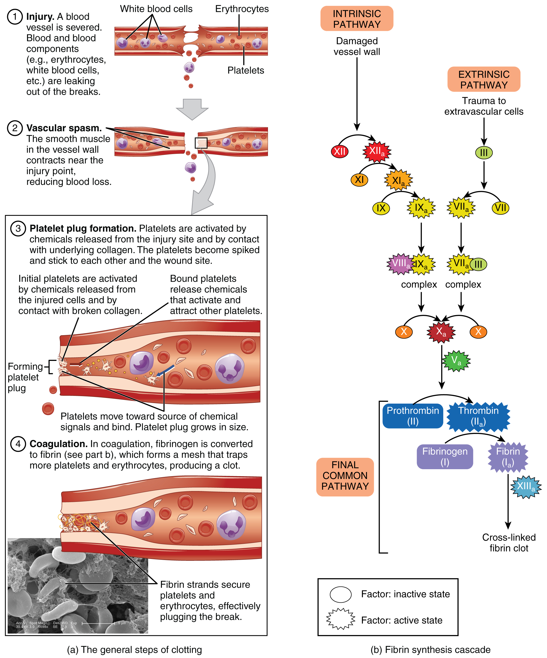
Hemostasis · Anatomy and Physiology
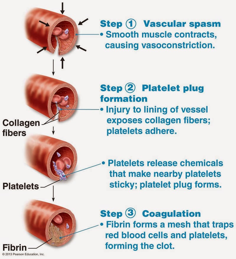
Bio Geo Nerd Blood Clotting

The Clotting Cascade Made Easy! Nurse Your Own Way
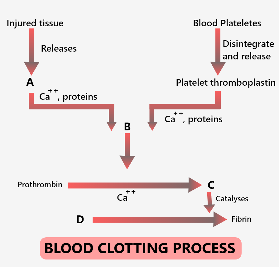
In the extrinsic clotting pathway, the active factor VII activates
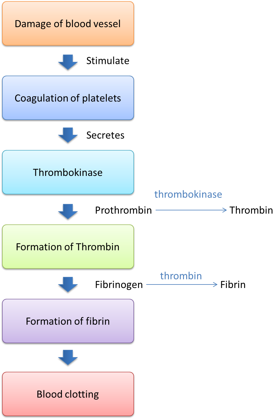
1.3.1 Mechanism of Blood Clotting SPM Biology

Flow Chart Blood Clotting Process Diagram

Blood clotting (Edexcel Alevel Biology B) Teaching Resources

Blood Clotting Cascade Diagram

Blood Clotting Cascade Diagram
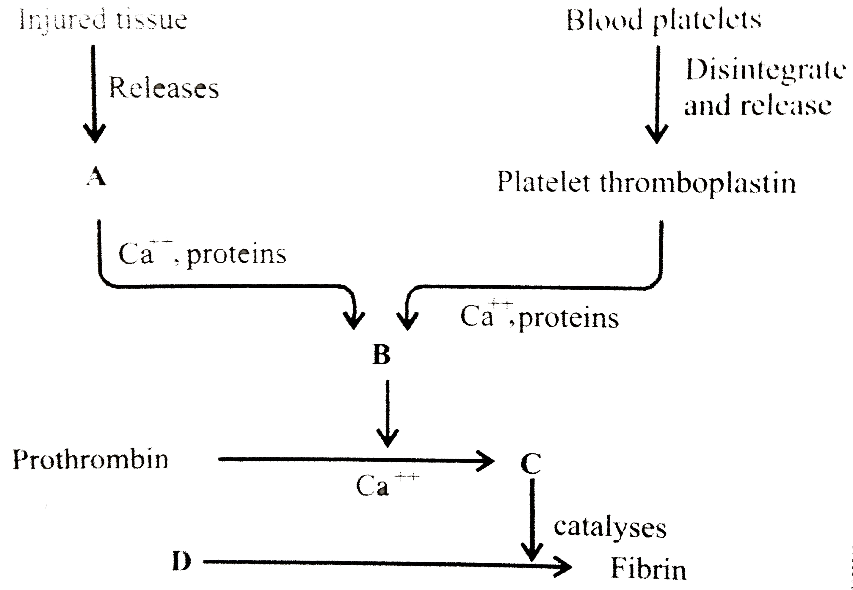
Describe the process of blood clotting.
The Process Of Blood Coagulation Leads To Haemostasis, I.e.
The Clot Will Dissolve After The Damage Heals.
Streiff, Md, Johns Hopkins University School Of Medicine.
Web These Problems Cause Brain Cells To Die.
Related Post: