Equinus Gait Pattern
Equinus Gait Pattern - Several approaches have been suggested for its correction, including pharmacological, surgical, and physical therapy (pt) interventions. Gait type description treatment / management type 1 true equinus this gait pattern is characterized by toe walking due to spasticity in the calf muscles which causes the Equinus plus recurvatum knee and extended hip. Web it is characterized by toe walking, otherwise known as equinus. “drop foot”, “true equinus”, “apparent equinus” “genu recurvatum”, “jump gait”, and “crouch gait”. Web the primary gait compensation pattern observed for simulated ankle equinus was increased knee flexion at initial contact. Web common gait deviations in cp can be grouped into the gait patterns of spastic hemiplegia (drop foot, equinus with different knee positions) and spastic diplegia (true equinus, jump, apparent equinus and crouch) to facilitate communication. Web equinus is a condition in which the upward bending motion of the ankle joint is limited. Equinus can occur in one or both feet. Web common gait deviations in cp can be grouped into the gait patterns of spastic hemiplegia (drop foot, equinus with different knee positions) and spastic diplegia (true equinus, jump, apparent equinus and crouch) to facilitate communication. Equinus plus recurvatum knee and extended hip. Web watch this short video simulating gait with an equinus deformity. True equinus refers to a gait characterized by plantarflexion of the foot and ankle with respect to the leg and may be seen in stance and/or the swing phase of gait. The wgh classification system mainly focuses on pathologies of the ankle. Equinus can occur in one or both feet. Web type 2 has an equinus gait pattern but with spastic or contracted plantar flexors, which overpower an active dorsiflexor. Web failure to recognize apparent equinus is the most common error in observational gait analysis and is of more than academic importance. Web equinus is a condition in which the upward bending. Web it is characterized by toe walking, otherwise known as equinus. Web watch this short video simulating gait with an equinus deformity. The wgh classification system mainly focuses on pathologies of the ankle joint and. Gait type description treatment / management type 1 true equinus this gait pattern is characterized by toe walking due to spasticity in the calf muscles. Type 3 includes the ankle position of type 2, further adding abnormal function of the knee joint. It is caused by spasticity or contracture in the calf muscle and achilles tendon. Web type 2 has an equinus gait pattern but with spastic or contracted plantar flexors, which overpower an active dorsiflexor. Web the sagittal gait pattern should be identified and. A significant degree of knee flexion occurred ranging from 7° to 22° ( p = 0.001). Web watch this short video simulating gait with an equinus deformity. Equinus plus neutral knee and extended hip. Web failure to recognize apparent equinus is the most common error in observational gait analysis and is of more than academic importance. Web gait of spastic. It is caused by spasticity or contracture in the calf muscle and achilles tendon. Web the sagittal gait pattern should be identified and described, as the ankle and knee levels are linked by coupling (e.g., the plantar flexion knee extension couple), and treatment should therefore be indicated with caution. We included the baumann and strayer procedures, as well as the. Gait type description treatment / management type 1 true equinus this gait pattern is characterized by toe walking due to spasticity in the calf muscles which causes the If unilateral, causes include peroneal nerve palsy and l5 radiculopathy. Equinus plus neutral knee and extended hip. Web one of the aims of orthotic management is to produce a more normal gait. It is caused by spasticity or contracture in the calf muscle and achilles tendon. Web as with hemiplegic cerebral palsy, there are also four commonly observed types of gait patterns which impact a person’s walking ability. Equinus can occur in one or both feet. Gait type description treatment / management type 1 true equinus this gait pattern is characterized by. Web gait of spastic mice was characterized by a typical hopping pattern. Web the primary gait compensation pattern observed for simulated ankle equinus was increased knee flexion at initial contact. Equinus plus neutral knee and extended hip. The movement of the skeletal foot allows you to view the foot from different perspectives. Web we found that improvements in the equinus. Equinus plus neutral knee and extended hip. Web the primary gait compensation pattern observed for simulated ankle equinus was increased knee flexion at initial contact. Web common gait deviations in cp can be grouped into the gait patterns of spastic hemiplegia (drop foot, equinus with different knee positions) and spastic diplegia (true equinus, jump, apparent equinus and crouch) to facilitate. Web one of the aims of orthotic management is to produce a more normal gait pattern by positioning joints in the proper position to reduce pathological reflex or spasticity. Type 3 includes the ankle position of type 2, further adding abnormal function of the knee joint. Web we found that improvements in the equinus gait and increases in the flexion angle of the swing phase in the hip joints resulted in increased walking speed, extended stride, and improved. Equinus plus recurvatum knee and extended hip. Equinus can occur in one or both feet. Web the sagittal gait pattern should be identified and described, as the ankle and knee levels are linked by coupling (e.g., the plantar flexion knee extension couple), and treatment should therefore be indicated with caution. True equinus refers to a gait characterized by plantarflexion of the foot and ankle with respect to the leg and may be seen in stance and/or the swing phase of gait. Someone with equinus lacks the flexibility to bring the top of the foot toward the front of the leg. Web as with hemiplegic cerebral palsy, there are also four commonly observed types of gait patterns which impact a person’s walking ability. Web equinus deformity of the foot is a common feature of hemiplegia, which impairs the gait pattern of patients. Web common gait deviations in cp can be grouped into the gait patterns of spastic hemiplegia (drop foot, equinus with different knee positions) and spastic diplegia (true equinus, jump, apparent equinus and crouch) to facilitate communication. (steppage gait, equine gait) seen in patients with foot drop (weakness of foot dorsiflexion), the cause of this gait is due to an attempt to lift the leg high enough during walking so that the foot does not drag on the floor. Web type 2 has an equinus gait pattern but with spastic or contracted plantar flexors, which overpower an active dorsiflexor. Web the primary gait compensation pattern observed for simulated ankle equinus was increased knee flexion at initial contact. Web this study aimed to evaluate gait outcomes and strength following the surgical correction of equinus in cerebral palsy (cp) based on different surgical procedures. A significant degree of knee flexion occurred ranging from 7° to 22° ( p = 0.001).
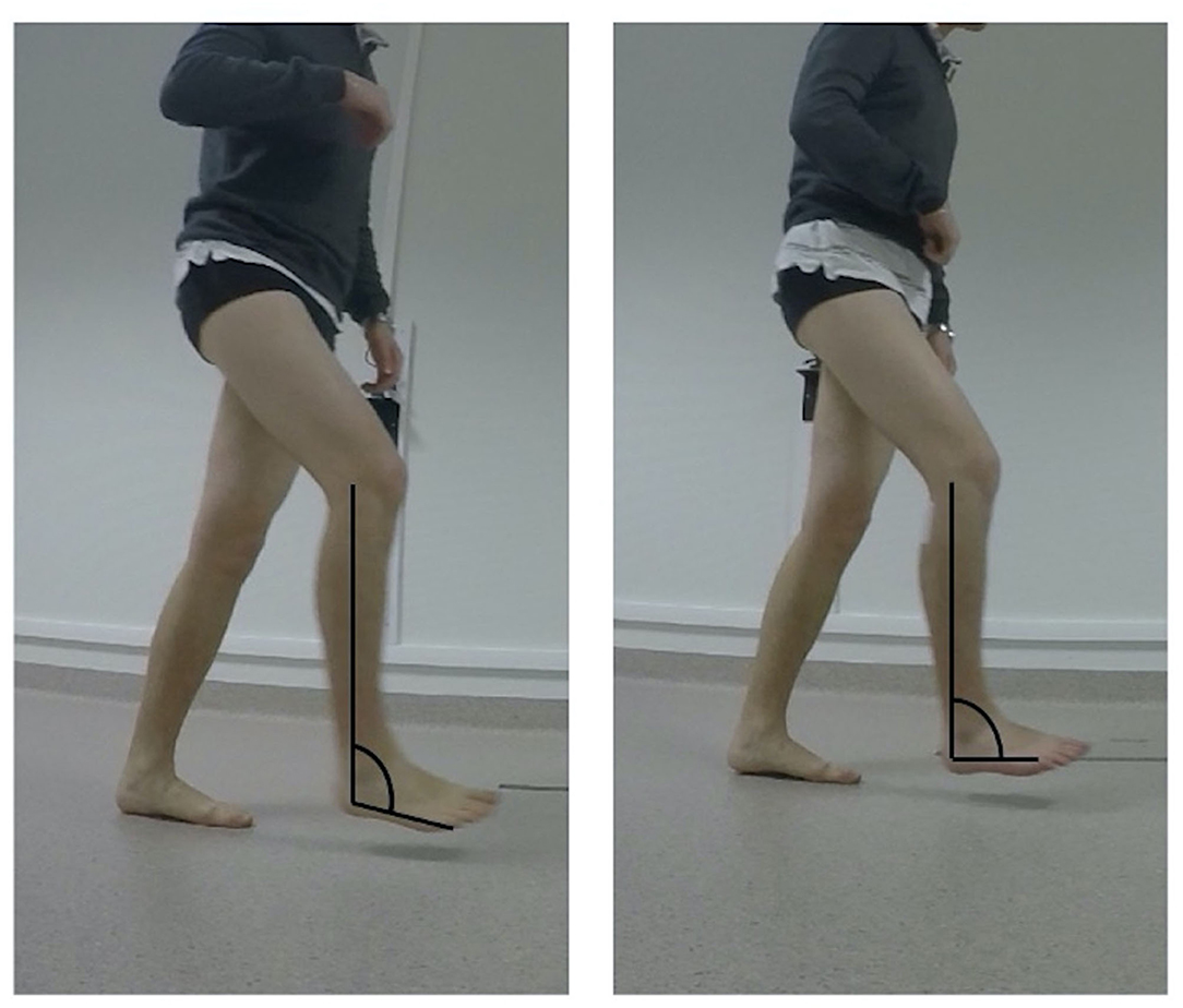
Lateral view photograph during gradual equinus deformity correction

Frontiers Respective Contributions of Instrumented 3D Gait Analysis

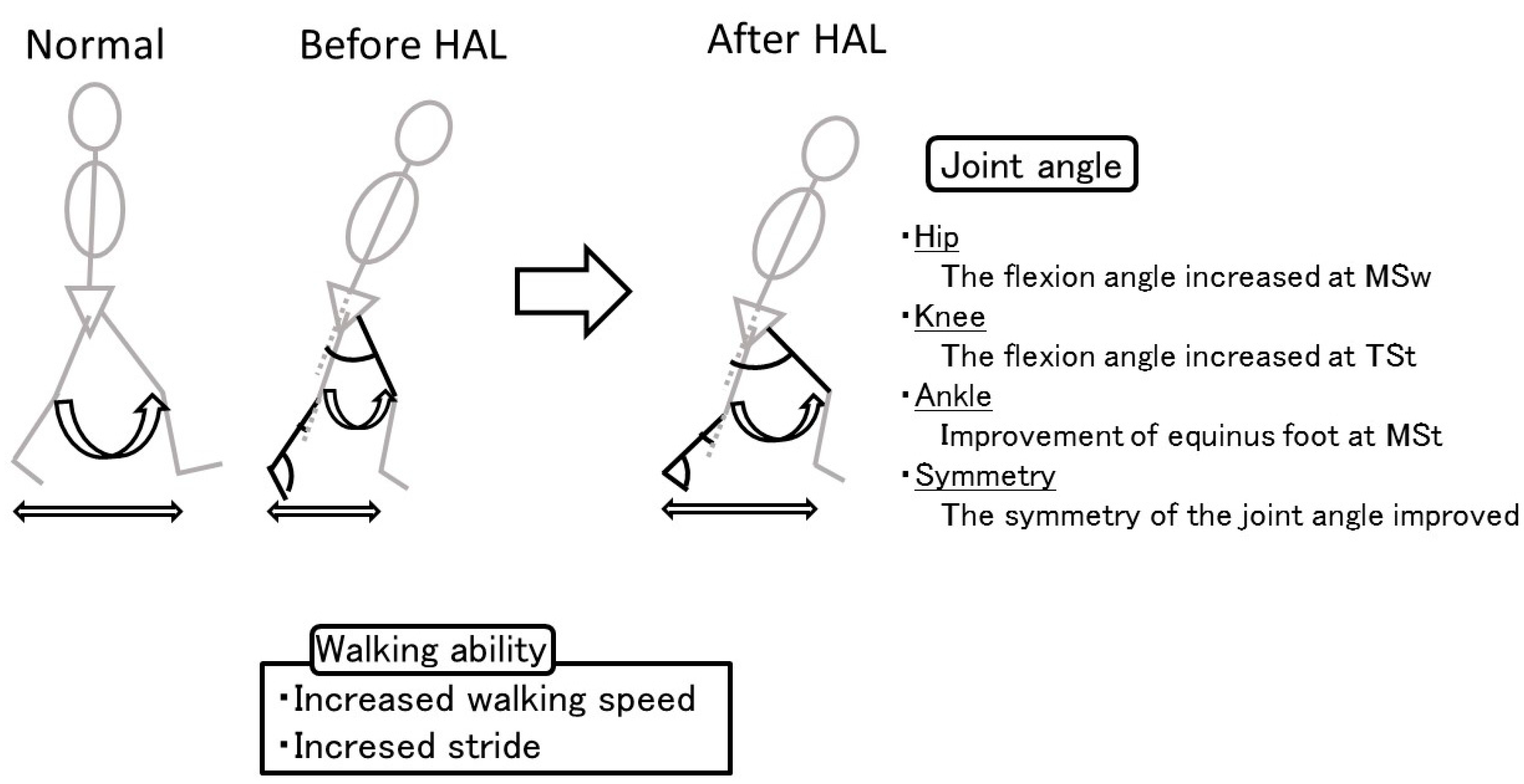
Gait pattern. The improvements in equinus gait and flexion angle of the

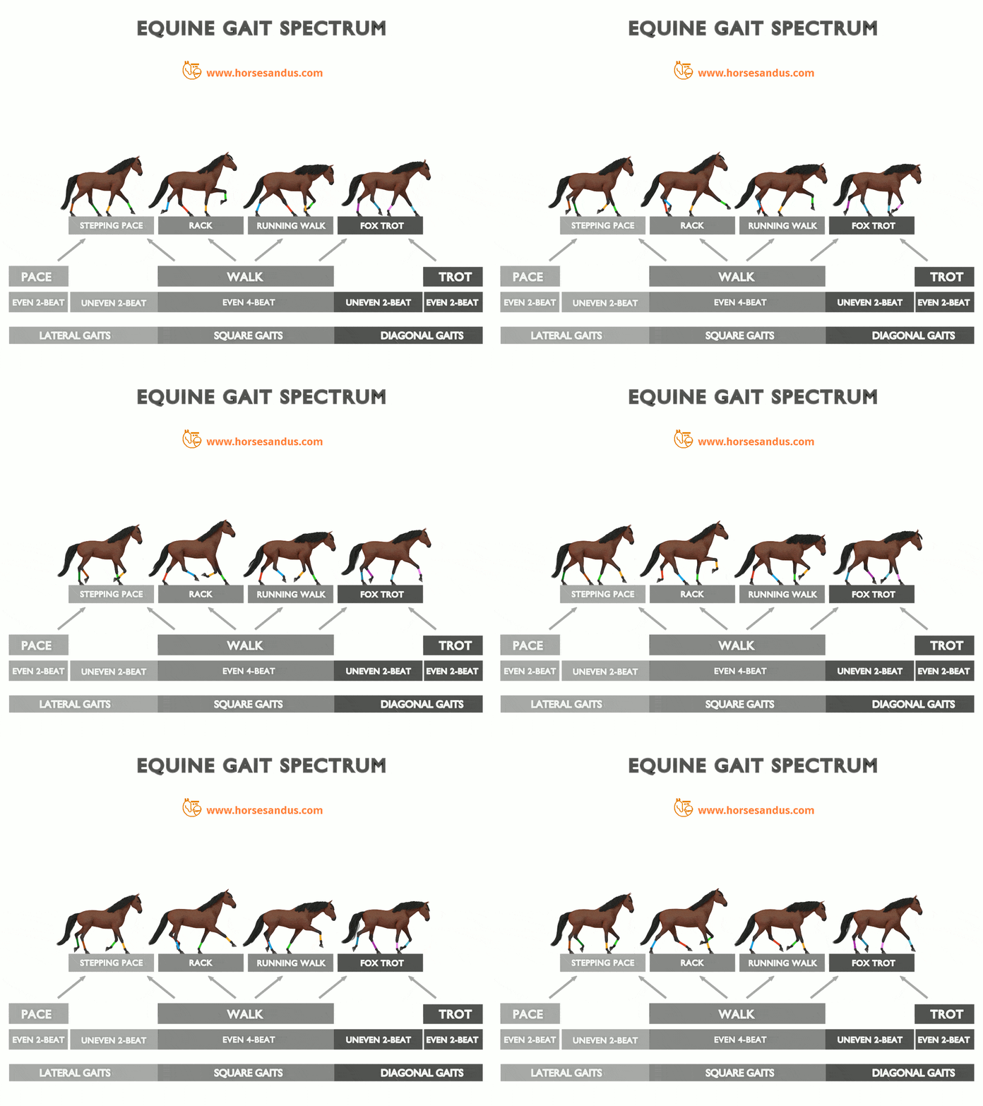
The “Ambling” Horse Gaits Complete Guide Horses and Us

Figure 1 from Multilevel surgery for equinus gait in children with

Gait pattern. The improvements in equinus gait and flexion angle of the

Medicina Free FullText Effect of the Hybrid Assistive Limb on the

Gait pattern. The improvements in equinus gait and flexion angle of the

*Gait 2* Physical Therapist Assistant 180 with P. Hill at Washtenaw

Gait pattern classification in children with cp
Web Equinus (Toe Walking) Gait This Type Of Gait Is Usually Observed In Children With Clubfoot Deformity (Equinovarus).
Gait Type Description Treatment / Management Type 1 True Equinus This Gait Pattern Is Characterized By Toe Walking Due To Spasticity In The Calf Muscles Which Causes The
Web In A True Equinus Gait Pattern, Calf Spasticity Or Fixed Ankle Equinus Contracture Is Dominant, Resulting In Excess Ankle Pf With Hips And Knees Either Extended Or Displaying Relatively Little Deviation From Normal Over The Gait Cycle.
If Unilateral, Causes Include Peroneal Nerve Palsy And L5 Radiculopathy.
Related Post: