Ecg Pattern In Pulmonary Embolism
Ecg Pattern In Pulmonary Embolism - Web background pulmonary embolism is a common and potentially fatal condition. Dvt/pe symptomsdvt/pe risk factorsdvt/pe facts Web pulmonary embolism can produce a wide variety of ecg changes. Web sinus tachycardia is the most common ecg finding in pulmonary embolism. Web tachycardia is the most common ecg finding (se ecg in pulmonary embolism below). Web there is a wide range of ecg features associated with pe. Web ecg changes with pulmonary embolism. Web unique ecg findings in acute pulmonary embolism: Exogenous estrogens in contraceptives are associated with an increased risk of venous. Web utilization of the electrocardiogram (ecg) to assess the diagnostic probability and severity of pulmonary embolism (pe) remains common in clinical. Web sinus tachycardia is the most common ecg finding in pulmonary embolism. Web there is a wide range of ecg features associated with pe. Pulmonary embolism (pe) is the third most common cause of death from cardiovascular disease. Web the ecg changes associated with acute pulmonary embolism may be seen in any condition that causes acute pulmonary hypertension, including hypoxia. Safety informationdoctor discussion guidecost informationtalking to your doctor Web there is a wide range of ecg features associated with pe. Pe occurs when a deep vein thrombosis. Distended jugular vein (due to elevated right ventricular pressure). Pulmonary embolism (pte, pe) ranges from asymptomatic to a life threatening catastrophe. Web a variety of electrocardiographic (ecg) changes have been thought to have diagnostic value in patients with suspected pulmonary embolism (pe), but most. Dvt/pe symptomsdvt/pe risk factorsdvt/pe facts Pe occurs when a deep vein thrombosis. Web ecg changes with pulmonary embolism. Pulmonary embolism (pte, pe) ranges from asymptomatic to a life threatening catastrophe. Pe occurs when a deep vein thrombosis. Web there is a wide range of ecg features associated with pe. Pulmonary embolism (pe) is the third most common cause of death from cardiovascular disease. Safety informationdoctor discussion guidecost informationtalking to your doctor Web a variety of electrocardiographic (ecg) changes have been thought to have diagnostic value in patients with suspected pulmonary. Tbe anterior subepicardial ischemic pattern is the most frequent ecg sign of massive pe. Web sinus tachycardia is the most common ecg finding in pulmonary embolism. Web the ecg changes associated with acute pulmonary embolism may be seen in any condition that causes acute pulmonary hypertension, including hypoxia causing pulmonary hypoxic vasoconstriction. The s1q3t3 pattern is a classic. Exogenous estrogens. Web pulmonary embolism can produce a wide variety of ecg changes. Pulmonary embolism (pe) is the third most common cause of death from cardiovascular disease. Web tachycardia is the most common ecg finding (se ecg in pulmonary embolism below). Safety informationdoctor discussion guidecost informationtalking to your doctor The most common ecg finding in pe is sinus tachycardia. The most common ecg finding in pe is sinus tachycardia. Web there is a wide range of ecg features associated with pe. Pe occurs when a deep vein thrombosis. The s1q3t3 pattern is a classic. Web sinus tachycardia is the most common ecg finding in pulmonary embolism. Web ecg changes with pulmonary embolism. Exogenous estrogens in contraceptives are associated with an increased risk of venous. Safety informationdoctor discussion guidecost informationtalking to your doctor Web utilization of the electrocardiogram (ecg) to assess the diagnostic probability and severity of pulmonary embolism (pe) remains common in clinical. Web interest in diagnosis of acute pulmonary embolism (pe) utilizing the electrocardiogram (ecg). The most common ecg finding in pe is sinus tachycardia. Dvt/pe symptomsdvt/pe risk factorsdvt/pe facts Pulmonary embolism (pte, pe) ranges from asymptomatic to a life threatening catastrophe. Rv dysfunction, pulmonary embolism, digital biomarkers, ecg analysis application, emergency department. Web background pulmonary embolism is a common and potentially fatal condition. Exogenous estrogens in contraceptives are associated with an increased risk of venous. Distended jugular vein (due to elevated right ventricular pressure). Web pulmonary embolism can produce a wide variety of ecg changes. Web tachycardia is the most common ecg finding (se ecg in pulmonary embolism below). Web sinus tachycardia is the most common ecg finding in pulmonary embolism. Web pulmonary embolism can produce a wide variety of ecg changes. The s1q3t3 pattern is a classic. Web ecg changes with pulmonary embolism. Exogenous estrogens in contraceptives are associated with an increased risk of venous. Dvt/pe symptomsdvt/pe risk factorsdvt/pe facts Web interest in diagnosis of acute pulmonary embolism (pe) utilizing the electrocardiogram (ecg) has decreased since the creation of imaging techniques such. Safety informationdoctor discussion guidecost informationtalking to your doctor Web tachycardia is the most common ecg finding (se ecg in pulmonary embolism below). Pe occurs when a deep vein thrombosis. Tbe anterior subepicardial ischemic pattern is the most frequent ecg sign of massive pe. Web unique ecg findings in acute pulmonary embolism: Web an electrocardiogram, or ecg/ekg, records the electrical activity of the heart to diagnose conditions such as pulmonary embolism. Web background pulmonary embolism is a common and potentially fatal condition. Safety informationdoctor discussion guidecost informationtalking to your doctor Web sinus tachycardia is the most common ecg finding in pulmonary embolism. This parameter is easy to obtain and reflects the severity of pe.
S1Q3T3 pattern on ECG in pulmonary embolism

Pulmonary Embolism Ecg

Pulmonary Embolism (PE) Causes, symptoms, diagnosis, treatment
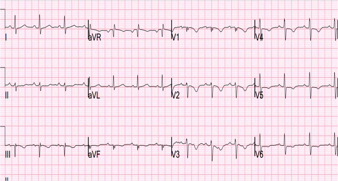
Pulmonary embolism ECG case study
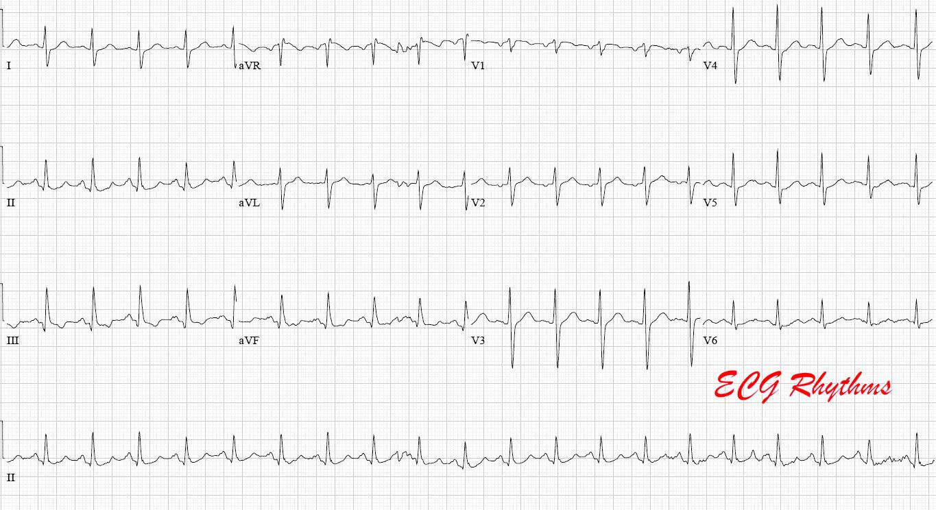
ECG Rhythms Pulmonary Embolism
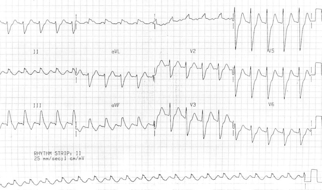
ECG changes in Pulmonary Embolism • LITFL • ECG Library
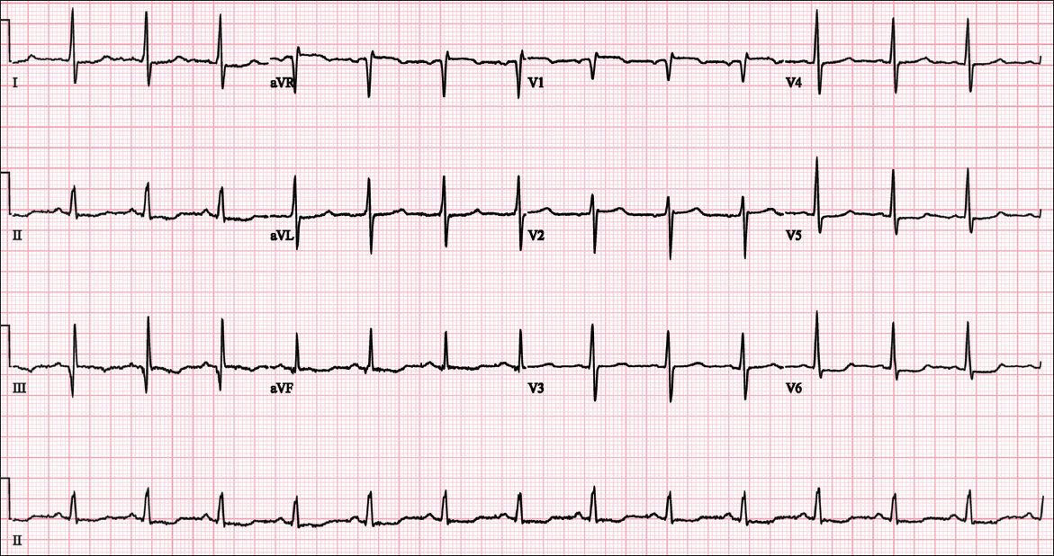
Electrocardiographic findings in pulmonary embolism SMJ
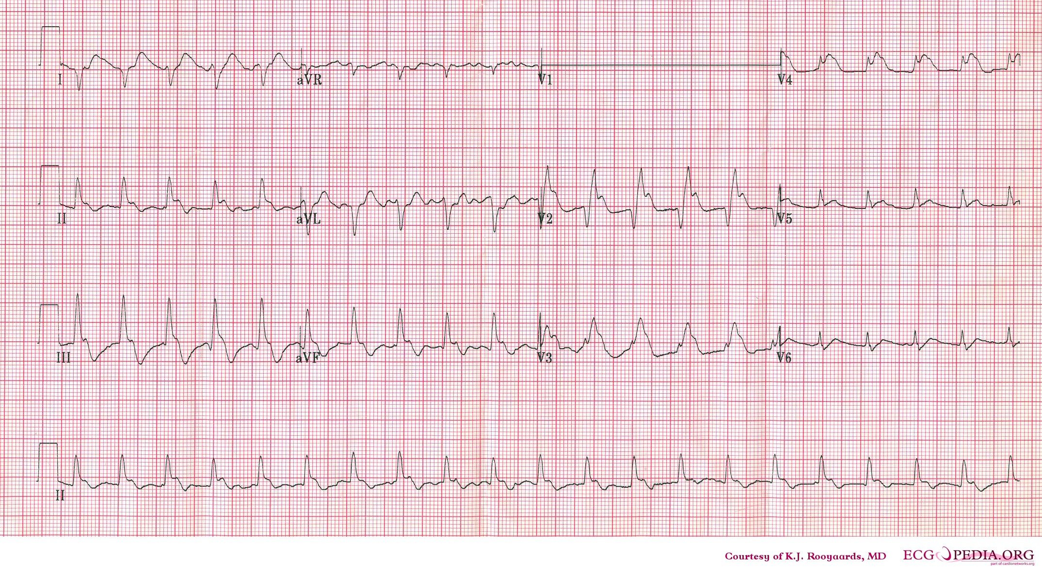
Pulmonary embolism electrocardiogram wikidoc

The ECG's of Pulmonary Embolism Resus
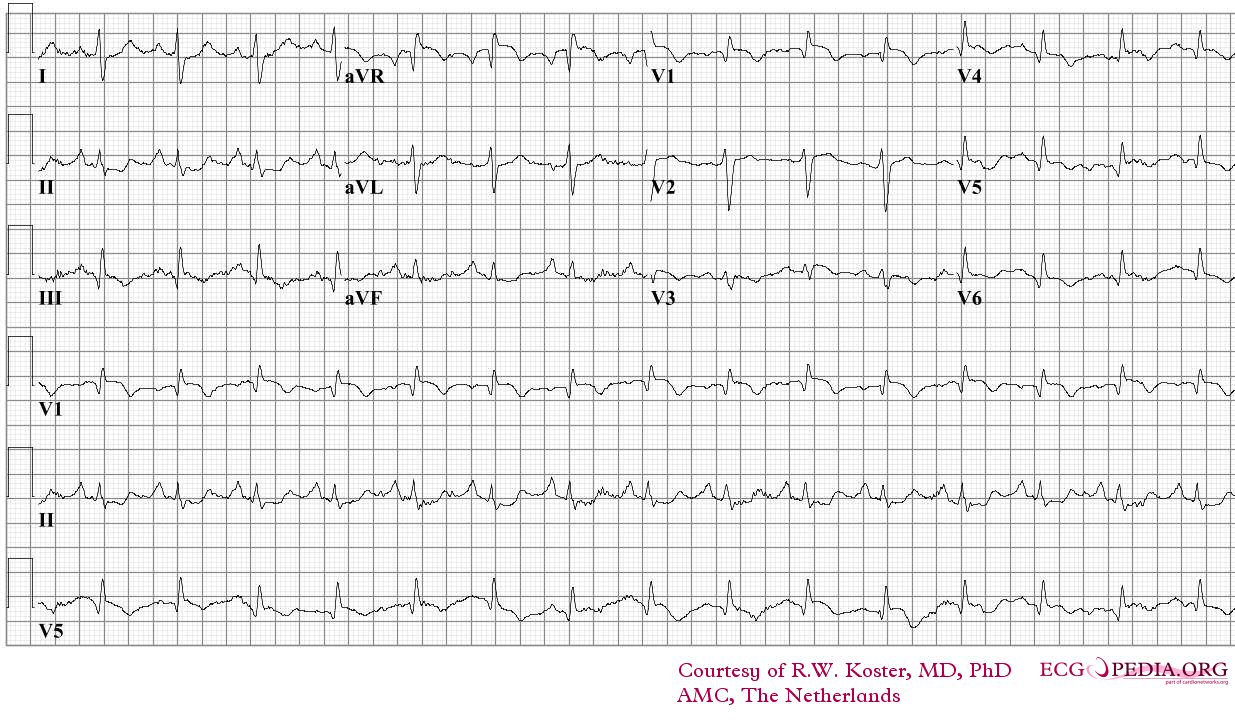
Pulmonary embolism electrocardiogram wikidoc
Web The Ecg Changes Associated With Acute Pulmonary Embolism May Be Seen In Any Condition That Causes Acute Pulmonary Hypertension, Including Hypoxia Causing Pulmonary Hypoxic Vasoconstriction.
Web A Variety Of Electrocardiographic (Ecg) Changes Have Been Thought To Have Diagnostic Value In Patients With Suspected Pulmonary Embolism (Pe), But Most.
Web Sinus Tachycardia Is The Most Common Ecg Finding In Pulmonary Embolism.
The Most Common Ecg Finding In Pe Is Sinus Tachycardia.
Related Post: