Dinoflagellates Drawing
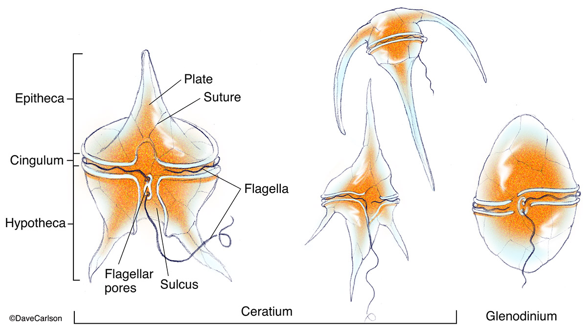
Dinoflagellates Drawing - Study the structure of dinoflagellate carefully. Web by integrating dinoflagellate molecular, fossil, and biogeochemical evidence, we propose a revised model for the evolution of thecal tabulations and suggest that the late acquisition. Web how to draw dinoflagellates Roughly half of the species in the group are photosynthetic (gaines and elbrächter 1987), the other half. Cells are covered by a theca (sheath) that can be smooth. Ceratium hirundinella, peridinium globulus) and nonthecate (e.g. Draw a dinoflagellate diagram and label it accurately. This section describes the life history and ecology of dinoflagellates, and. Their size is typically between 20 and 350 μm, rarely more or less. Web dinoflagellates are a group of unicellular organisms characterized by the possession of two flagella, one of which is directed transversely and the other longitudinally. They have 2 unlike flagella cropping up from the side of the ventral cell (dinokont flagellation). Dinoflagellate drawing stock photos are. Cells are covered by a theca (sheath) that can be smooth. Dinoflagellates are some of the most common eukaryotic cells in the ocean, but have very unusual nuclei. Many exhibit a form of closed. A guide to culturing algae. Web dinoflagellates are the most common sources of bioluminescence at the surface of the ocean. Dinoflagellates are some of the most common eukaryotic cells in the ocean, but have very unusual nuclei. Web dinoflagellates are common organisms in all types of aquatic ecosystems. Dinoflagellate drawing stock photos are. Predominant colour is golden brown but yellow, green, brown. Many exhibit a form of closed. Web the cell wall or theca of many motile dinoflagellates is armored and consists of cellulose plates that form a distinctive geometry or tabulation, whereas the cysts only bear traces of the theca tabulation. Web dinoflagellates are common organisms in all types of aquatic ecosystems.. Web the cell wall or theca of many motile dinoflagellates is armored and consists of cellulose plates that form a distinctive geometry or tabulation, whereas the cysts only bear traces of the theca tabulation. Web dinoflagellates are the most common sources of bioluminescence at the surface of the ocean. B, epitheca (s is a sulcal plate); Their size is typically. Web dinoflagellates are the most common sources of bioluminescence at the surface of the ocean. A guide to culturing algae. Web (i) din flagellates are basically unicellular motile and biflagellate, golden brown, photosynthetic protists. (b) gonyaulax spinifera a, ventral; Dinoflagellates are found in fresh and saltwater. Web instructor derrick arrington view bio. Dinoflagellates are found in fresh and saltwater. Ceratium hirundinella, peridinium globulus) and nonthecate (e.g. A guide to culturing algae. It is known to cause bioluminescence in the ocean. Learn about the characteristics of dinoflagellates, how they move, and what they are best described as. Web dinoflagellates are the most common sources of bioluminescence at the surface of the ocean. Cells are covered by a theca (sheath) that can be smooth. Web the cell wall or theca of many motile dinoflagellates is armored and consists of cellulose plates that. A guide to culturing algae. Web by integrating dinoflagellate molecular, fossil, and biogeochemical evidence, we propose a revised model for the evolution of thecal tabulations and suggest that the late acquisition. Cells are covered by a theca (sheath) that can be smooth. Web (i) din flagellates are basically unicellular motile and biflagellate, golden brown, photosynthetic protists. Roughly half of the. Web several dinoflagellates, both thecate (e.g. Predominant colour is golden brown but yellow, green, brown. And kofoidinium spp.), draw prey to. Web how to draw dinoflagellates Web drawing on the advances in knowledge of both the phylogenetic history of dinoflagellates and the biochemical properties of their nuclei, we now are able to hypothesize the. Web dinoflagellates are a group of unicellular organisms characterized by the possession of two flagella, one of which is directed transversely and the other longitudinally. This section describes the life history and ecology of dinoflagellates, and. Web how to draw dinoflagellates Predominant colour is golden brown but yellow, green, brown. (b) gonyaulax spinifera a, ventral; Dinoflagellates are found in fresh and saltwater. Web dinoflagellates are common organisms in all types of aquatic ecosystems. Dinoflagellates are some of the most common eukaryotic cells in the ocean, but have very unusual nuclei. Web by integrating dinoflagellate molecular, fossil, and biogeochemical evidence, we propose a revised model for the evolution of thecal tabulations and suggest that the late acquisition. Study the structure of dinoflagellate carefully. A guide to culturing algae. Many exhibit a form of closed. Their size is typically between 20 and 350 μm, rarely more or less. Web the cell wall or theca of many motile dinoflagellates is armored and consists of cellulose plates that form a distinctive geometry or tabulation, whereas the cysts only bear traces of the theca tabulation. Cells are covered by a theca (sheath) that can be smooth. Web line drawings of some armored dinoflagellates. Web drawing on the advances in knowledge of both the phylogenetic history of dinoflagellates and the biochemical properties of their nuclei, we now are able to hypothesize the. This section describes the life history and ecology of dinoflagellates, and. And kofoidinium spp.), draw prey to. Web several dinoflagellates, both thecate (e.g. Dinoflagellate drawing stock photos are.
Dinoflagellate ceratium longpipe High School Science Teacher, High

Exploring dinoflagellate biology with the awesome power of yeast

Dinoflagellate Marine, Microscopic, Plankton Britannica

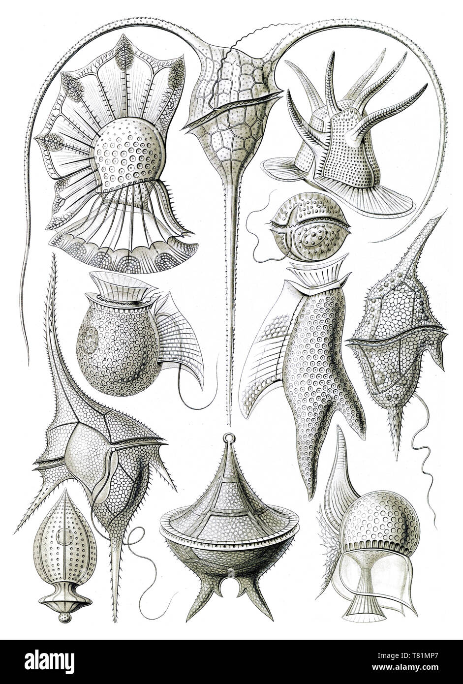
Various species of dinoflagellates armoured with cellulose plates drawn
Dinoflagellates ClipArt ETC

What is a Dinoflagellate? (with pictures)

Dinoflagellate Everything You Need to Know with Photos Videos

Ernst Haeckel, Dinoflagellates Stock Photo Alamy

Dinoflagellates Carlson Stock Art

NOAA_dinoflagellate_plankton Sea creatures art, Scientific
Web Instructor Derrick Arrington View Bio.
Roughly Half Of The Species In The Group Are Photosynthetic (Gaines And Elbrächter 1987), The Other Half.
Web Dinoflagellates Are The Most Common Sources Of Bioluminescence At The Surface Of The Ocean.
They Have 2 Unlike Flagella Cropping Up From The Side Of The Ventral Cell (Dinokont Flagellation).
Related Post: