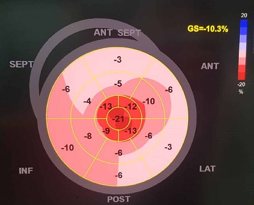
Amyloid Strain Pattern
Amyloid Strain Pattern - (a) thickening of the basal segments (solid lines) due to amyloid infiltration; Unfortunately, the diagnosis of ca is often made late and when the disease process is advanced. Web an apical sparing longitudinal strain pattern is typical of ca, wherein the apical strain is greater than two times the basal strain segments. 65% male, 62.5% amyloidosis light chain [al] type), 40 patients with hypertrophic cardiomyopathy. However, advances in cardiovascular imaging have allowed for better prognostication and establishing. Also studied regional function in ca patients. Web finally, amyloid infiltration impairs (c) global longitudinal strain (gls) characteristically with apical sparing of the lv apex, in contrast to a normal pattern, and severely reduced contractile function of the atrial myocardium, in contrast to. Right ventricular (rv) peak systolic strain. Lge was present in all patients with biopsy confirmed disease. When plotted on a bullseye, this will generate a characteristic “apical sparing” pattern visually. 2 the authors report that longitudinal strain (ls) of the left ventricle provides important information about the. Web cardiac amyloidosis (ca) results from cardiac infiltration with systemic light chains (al) or the transthyretin amyloid (attr) protein. Right ventricular (rv) peak systolic strain. Web in this issue of the european heart journal, cohen et al. From visual inspection, general trends can. Also studied regional function in ca patients. Global longitudinal strain (gls) is reduced and associated with survival in both al and attr. However, it is unclear how frequently this strain pattern truly. Cardiac involvement, namely cardiac amyloidosis (ca), occurs in up to 50 % of patients with primary amyloidosis and indicates almost invariably a grave prognosis. Web secondly, as strain. Web an apical sparing longitudinal strain pattern is typical of ca, wherein the apical strain is greater than two times the basal strain segments. Report the results of a detailed characterization of echocardiographic and haematology findings in 915 patients with al amyloidosis [69% of whom had cardiac amyloidosis (ca)]. Also studied regional function in ca patients. 2 the authors report. Unfortunately, the diagnosis of ca is often made late and when the disease process is advanced. Web regional longitudinal strain values (relative apical sparing or septal apical to base longitudinal strain ratio). Web four‐chamber strain imaging using speckle‐tracking echocardiography in a patient with biopsy‐verified light‐chain amyloidosis. (left) and strain pattern (centre and right) characteristic of an infiltrative process. In cardiac. Web a specific pattern of longitudinal strain characterized by worse longitudinal strain in the mid and basal ventricle with relative sparing of the apex 4 may help distinguish lv infiltration because of amyloid from true ventricular hypertrophy of hypertensive heart disease or hypertrophic cardiomyopathy. Web average strain pattern motifs. However, advances in cardiovascular imaging have allowed for better prognostication and. Web despite the extensive literature on the apical sparing of longitudinal strain in amyloidosis 36,52,54,. Web an apical sparing longitudinal strain pattern is typical of ca, wherein the apical strain is greater than two times the basal strain segments. Web average strain pattern motifs. There was significantly decreased global longitudinal strain and strain rate in the epicardial and endocardial layers.. Web figure 10 atypical strain pattern in a patient with prior anterior septal apical myocardial infarction and subsequent amyloid infiltration in the noninfarcted segments. Similar phasic strain graphs of the right atrium (ra). (a) thickening of the basal segments (solid lines) due to amyloid infiltration; There was significantly decreased global longitudinal strain and strain rate in the epicardial and endocardial. Lge was present in all patients with biopsy confirmed disease. Web a specific pattern of longitudinal strain characterized by worse longitudinal strain in the mid and basal ventricle with relative sparing of the apex 4 may help distinguish lv infiltration because of amyloid from true ventricular hypertrophy of hypertensive heart disease or hypertrophic cardiomyopathy. In cardiac amyloidosis the segmental strain. 2 the authors report that longitudinal strain (ls) of the left ventricle provides important information about the. The arrow demonstrates scarred segment from previous myocardial infarct. Web four‐chamber strain imaging using speckle‐tracking echocardiography in a patient with biopsy‐verified light‐chain amyloidosis. Atrial (la) strain showing reservoir and booster components. Lge was present in all patients with biopsy confirmed disease. In addition, its diagnostic value is not dependent on the underlying amyloidosis type. 15 amyloid infiltration was seen to cause greatly reduced longitudinal strain in all basal and mid regions, with a relative sparing of the lv apex. Web may 6, 2024 updated 12:19 p.m. Report the results of a detailed characterization of echocardiographic and haematology findings in 915 patients. Results of receiver‐operating characteristic analysis for the discrimination of cardiac amyloidosis by echocardiographic strain parameters. The arrow demonstrates scarred segment from previous myocardial infarct. Unfortunately, the diagnosis of ca is often made late and when the disease process is advanced. Atrial (la) strain showing reservoir and booster components. Web secondly, as strain imaging can measure regional myocardial deformation, phelan et al. Web despite the extensive literature on the apical sparing of longitudinal strain in amyloidosis 36,52,54,. Web the accuracy of an apical‐sparing strain pattern on transthoracic echocardiography (tte) for predicting cardiac amyloidosis (ca) has varied in prior studies depending on the underlying cohort. Web regional longitudinal strain values (relative apical sparing or septal apical to base longitudinal strain ratio). Atrial (la) strain showing reservoir and booster components. The average strain patterns motifs for each cardiac condition are shown (fig. Ejection fraction strain ratio and the other deformation parameters have overcome the shortcomings of conventional echocardiographic indices, which in our. Web four‐chamber strain imaging using speckle‐tracking echocardiography in a patient with biopsy‐verified light‐chain amyloidosis. Web may 6, 2024 updated 12:19 p.m. There was significantly decreased global longitudinal strain and strain rate in the epicardial and endocardial layers. Right ventricular (rv) peak systolic strain. (a) thickening of the basal segments (solid lines) due to amyloid infiltration;
Echo Parameters for Differential Diagnosis in Cardiac Amyloidosis

Characteristic appearance of cardiac amyloidosis on echocardiography

Relative apical sparing of longitudinal strain using twodimensional
Amyloidosis American Academy of Ophthalmology

Cureus Role of Echocardiography in the Diagnosis of Light Chain

Echocardiographic features of cardiac amyloidosis. A Apical 4 chamber

Global and Regional Variations in Transthyretin Cardiac Amyloidosis A
![]()
(PDF) Relative apical sparing of longitudinal strain using two

Bullseye display of longitudinal strain analysis in a patient with

What Is Lv Strain Pattern Natural Resource Department
Mets Manager Carlos Mendoza Said Sunday That Senga, Who Threw Live.
Web An Apical Sparing Longitudinal Strain Pattern Is Typical Of Ca, Wherein The Apical Strain Is Greater Than Two Times The Basal Strain Segments.
Loid Deposits Are The Result Of (1) Abnormally Functioning Plasma Cells Or (2) Mutations Of The Transthyretin Protein (Expressed By Hepatocytes).
Web Figure 10 Atypical Strain Pattern In A Patient With Prior Anterior Septal Apical Myocardial Infarction And Subsequent Amyloid Infiltration In The Noninfarcted Segments.
Related Post: